

Peptide concentration is most reliably assessed by measuring its amino acid composition after hydrolysis, then separating and detecting the released amino acids. Amino acid analysis for peptide concentration commonly uses 6N HCl at 110°C for 24-96h in vacuo, followed by HPLC separation and UV or fluorometric detection. The result depends on how well each residue survives hydrolysis, so a single method may not recover every amino acid equally.
That matters when the reported concentration needs to reflect the peptide itself rather than an assumption about its composition. Amino acid analysis is used as a reference method for peptide quantification, but sensitive residues and sulfur-containing amino acids require particular care during sample preparation and interpretation.
This guide explains how derivatization chemistry affects detection, why hydrolysis conditions matter for tryptophan and other sensitive amino acids, and how sulfur amino acid recovery can influence calculated concentration. It also covers post-column detection and practical considerations for making peptide concentration results more representative and reproducible.
Key Takeaways
- Match derivatization chemistry to the detection method and the amino acids being measured to support reliable quantitation.
- Choose hydrolysis conditions with sensitive residues in mind, since recovery can vary substantially with the chemistry used.
- Account for methionine and cysteine recovery when interpreting peptide composition and concentration results.
- Understand how post-column derivatization and detection affect signal generation, then consider EuroLab Peptides’ Longevity & Vitality Research in the context of research focused on longevity.
Amino acid derivatization chemistries
Amino acid derivatization is the chemical conversion of amino acids into forms suitable for detection. After peptide hydrolysis, released amino acids are separated by HPLC and measured using ultraviolet or fluorometric detection. Derivatization supports measurement by producing a detectable signal. The selected chemistry must work with both the amino acid profile and the detector.
Pre-column derivatization occurs before chromatographic separation; post-column derivatization occurs after separation. The choice affects workflow and signal generation. In either approach, standards and samples need compatible treatment. Otherwise, differences in reaction or response can distort comparisons between peaks. Concentrated peptide solutions may be added directly to hydrolysis reagent when they dilute the final reagent by no more than about 5%. This preserves the intended hydrolysis conditions.
Peptide concentration is calculated by converting measured amino acid quantities to molar amounts, then dividing by the number of corresponding residues in the peptide sequence. Estimates based on multiple residues can be compared to identify inconsistent recovery. This makes amino acid analysis for peptide concentration dependent on knowing the sequence and selecting appropriate residues for calculation.
Accuracy and precision depend on calibration, peak measurement, hydrolysis recovery, and consistent derivatization. A residue that degrades during hydrolysis can produce a low estimate, even when detection is repeatable. Comparing several suitable residues can help reveal this bias. Amino acid analysis is used as a reference method for peptide quantification, but results also reflect the method’s recovery characteristics.
Commercial laboratory timing includes sample preparation, hydrolysis, chromatographic analysis, and reporting. The standard hydrolysis step uses 6N HCl at 110°C for 24-96h in vacuo; instrument and reporting time add to that interval.
Required sample amount and handling depend on peptide concentration, amino acid composition, and analytical sensitivity. Provide a representative, uncontaminated sample and sufficient material for the laboratory’s validated workflow. For direct addition to hydrolysis reagent, keep sample dilution within the approximately 5% limit. Preserve sample identity and avoid handling that could introduce contaminants or cause material loss.
Hydrolysis of sensitive amino acids
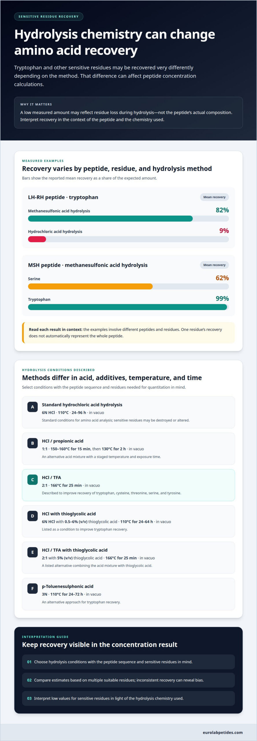
Hydrolysis breaks peptide bonds into amino acids. Standard hydrochloric acid conditions can destroy or alter sensitive residues. Tryptophan is especially vulnerable, so its measured recovery can vary substantially with the reagents used. This can bias concentration calculations based on amino acid composition.
Recovery is the measured amount of an amino acid relative to its expected amount. In LH-RH peptide, methanesulfonic acid hydrolysis produced mean tryptophan recovery of 82%. Hydrochloric acid hydrolysis produced 9%. For MSH peptide, methanesulfonic acid yielded 62% mean serine recovery and 99% mean tryptophan recovery. These examples show why one residue’s result cannot automatically represent the whole peptide.
Alternative acid mixtures can change both exposure time and recovery. One method uses HCl/Propionic acid (1:1) at 150 – 160 o C for 15 min, then 130 o C for 2h in vacuo. Another uses HCl/TFA (2:1) at 166ºC for 25 min in vacuo to improve recovery of tryptophan, cysteine, threonine, serine, and tyrosine.
Thioglycolic acid is an additive used to support recovery of sensitive residues. A listed condition combines 6 N HCl with 0.5-6% (v/v) thioglycolic acid at 110 o C for 24-64h in vacuo to improve tryptophan recovery. Another combines HCl/TFA (2:1) with 5% (v/v) thioglycolic acid at 166ºC for 25 min in vacuo. These conditions differ in acid composition, additive level, temperature, and duration.
p-Toluenesulphonic acid hydrolysis is an alternative approach for tryptophan recovery. The listed method uses 3N p-Toluenesulphonic acid at 110 o C for 24-72 h in vacuo. Select conditions according to the peptide’s sequence and the residues needed for quantitation. Compare recovery across suitable residues, and interpret low sensitive-residue values in light of the hydrolysis chemistry used.
Methionine and cysteine recovery
Sulfur amino acids are residues that contain sulfur, including methionine and cysteine. Their measured amounts can change during acid hydrolysis, complicating peptide composition calculations. Methionine may be altered by oxidation, while cysteine can form products that differ from the original residue. A chromatographic result therefore reflects both the peptide sequence and the chemistry used to prepare the sample.
Recovery is the amount of an amino acid measured after preparation relative to its amount in the sample. Low recovery of methionine or cysteine can make a peptide appear less concentrated when calculations rely on those residues. It can also skew residue mole percentages relative to the known peptide sequence. Interpret each sulfur-containing residue in light of its expected abundance and the selected hydrolysis conditions.
Sodium sulfite hydrolysis is a method used to improve recovery of methionine, cysteine, and tyrosine. The listed condition uses 6N HCL-Na₂SO₃ at 110 o C for 24h in vacuo. Sodium sulfite is included in the hydrolysis chemistry; the method’s purpose is to improve measurement of those residues. The listed condition includes all three amino acids, so it can be relevant when the peptide sequence contains them.
For concentration analysis, compare estimates based on different residues instead of relying on a single sulfur amino acid. First, identify the methionine and cysteine counts in the sequence. Then determine whether the method is designed to assess their recovery. Finally, compare their measured contributions with those of other suitable residues. Differences between estimates may indicate residue-specific recovery effects rather than a change in peptide amount.
Record the hydrolysis reagent and conditions alongside the reported amino acid values. For example, distinguish a result produced with 6N HCL-Na₂SO₃ at 110 o C for 24h in vacuo from one using another hydrolysis chemistry. That context supports reproducible interpretation and prevents direct comparisons between results generated under unlike conditions. For synthetic peptides, sequence-based residue percentages provide an additional comparison point, especially when sulfur residues contribute materially to the expected composition.
Post-column derivatization and detection
Post-column derivatization is a reaction performed after amino acids leave the separation column. A reagent reacts with each separated amino acid to produce a detectable signal. Because the reaction follows chromatography, it supports measurement of amino acids as distinct peaks. UV absorbance or fluorometry can then measure the resulting signal.
Pre-column UV detection uses derivatization before the sample enters the column. The reagent adds a UV-absorbing group to amino acids, allowing their separated peaks to be measured. In this setup, derivatization is part of sample preparation; post-column chemistry takes place in the detection flow path. The choice affects workflow and how reaction conditions are controlled.
Detection converts each peak into a measurable response. Calibration relates that response to known amino acid quantities, allowing sample results to be quantified. Standards and samples should undergo compatible preparation; otherwise, differences in derivatization can affect peak comparisons. Peak identity also matters: concentration calculations should use amino acids associated with the peptide, not unrelated signals.
Amino acid analysis measures individual amino acids released by hydrolysis, while UV absorbance estimates peptide amount from the sample’s absorbance. The UV estimate can depend on peptide composition and other absorbing material. BCA and Bradford assays are colorimetric methods that are easy to perform and require little instrumentation, but results can vary between samples and may be inaccurate.
For repeated measurements of the same protein, a colorimetric assay can be calibrated against a standard whose concentration was established by amino acid analysis. This links a convenient signal to a composition-based reference. The calibration is most useful when the protein stays consistent; changing the peptide or sample matrix can alter the assay response.
Choose a detection method based on the measurement goal. Use composition data when residue-level quantitation matters. Use UV or colorimetric assays for convenient estimates, while accounting for composition and sample effects. Document the derivatization step, calibration standard, and detection mode so results can be interpreted and compared accurately.
EuroLab Peptides: Longevity & Vitality Research
EuroLab Peptides offers Longevity & Vitality Research for scientific programs examining peptide applications in longevity-related research. The analytical methods covered above help researchers assess peptide composition and concentration, while this offering provides a relevant research focus, not an amino acid analysis service. It may suit work with research objectives centered on longevity and vitality. Other research areas include Weight Management Research, Tissue Repair & Recovery Research, and Cognitive Function Research. Match the offering to the study’s scientific focus, keeping analytical characterization distinct from research scope. For research focused on longevity and vitality, visit eurolabpetides.com to explore the relevant offering.
Align the Next Research Decision
Use amino acid analysis for peptide concentration to support a defined research decision, not as an isolated result. Before interpreting data, establish how the measured concentration will inform the study and which analytical assumptions matter. Recording these decisions can support consistent comparisons as experiments develop. The next step is to connect analytical planning with the research question the study is designed to address.
EuroLab Peptides’ Longevity & Vitality Research, Weight Management Research, and Tissue Repair & Recovery Research address distinct scientific research areas. Consider the area that best aligns with your project’s focus, while keeping peptide characterization separate from the broader research objective. Explore EuroLab Peptides’ research offerings as you plan the next stage of your work. A clearly framed research question gives each subsequent decision a stronger foundation.
Frequently Asked Questions
How is amino acid analysis used to determine peptide concentration?
Measured amino acid quantities are related to residue counts in the peptide sequence to estimate peptide amount. For example, if the sequence contains two copies of a measured residue, interpret its detected amount in relation to both copies. Convert the resulting molar peptide concentration to mass concentration using the peptide’s molecular mass. Comparing estimates based on sequence-compatible residues can reveal discrepancies.
What is the principle of amino acid analysis for peptide quantification?
The principle is compositional: peptide bonds are cleaved, amino acids are separated, and detector signals are compared with calibrated standards. The resulting profile gives quantities for individual residues that can be interpreted against the known sequence. This differs from measuring an intact molecule directly. The method is most informative when sequence identity is established independently and the selected residues have interpretable recovery.
What sample preparation and hydrolysis steps are required for peptide amino acid analysis?
Prepare a representative sample aliquot and link its identity to the peptide sequence and expected composition. Hydrolyze it under controlled acidic conditions, then prepare the resulting mixture for chromatographic analysis using the validated method. Include reagent blanks and amino acid standards to distinguish sample-derived peaks from background. Record the hydrolysis chemistry because it can affect residue recovery and the concentration calculation.
Which amino acid analysis methods are used for peptide concentration, such as HPLC or LC-MS?
HPLC separates released amino acids before UV or fluorometric measurement, often after derivatization to produce detectable signals. LC-MS detects analytes through mass-to-charge signals when the analytical method is designed for that purpose. These platforms are not interchangeable by name alone. When selecting a method for concentration analysis, compare validation, target analytes, calibration, and hydrolysis recovery.
Which amino acids are unstable or difficult to quantify after peptide hydrolysis?
Tryptophan is particularly vulnerable during acid treatment, while methionine and cysteine require careful recovery assessment. Serine, threonine, and tyrosine can also be affected by hydrolysis conditions. For peptides containing these residues, interpret results against the sequence and the selected method. Reporting the hydrolysis chemistry alongside residue measurements helps readers assess whether low values reflect sample composition or preparation effects.
