
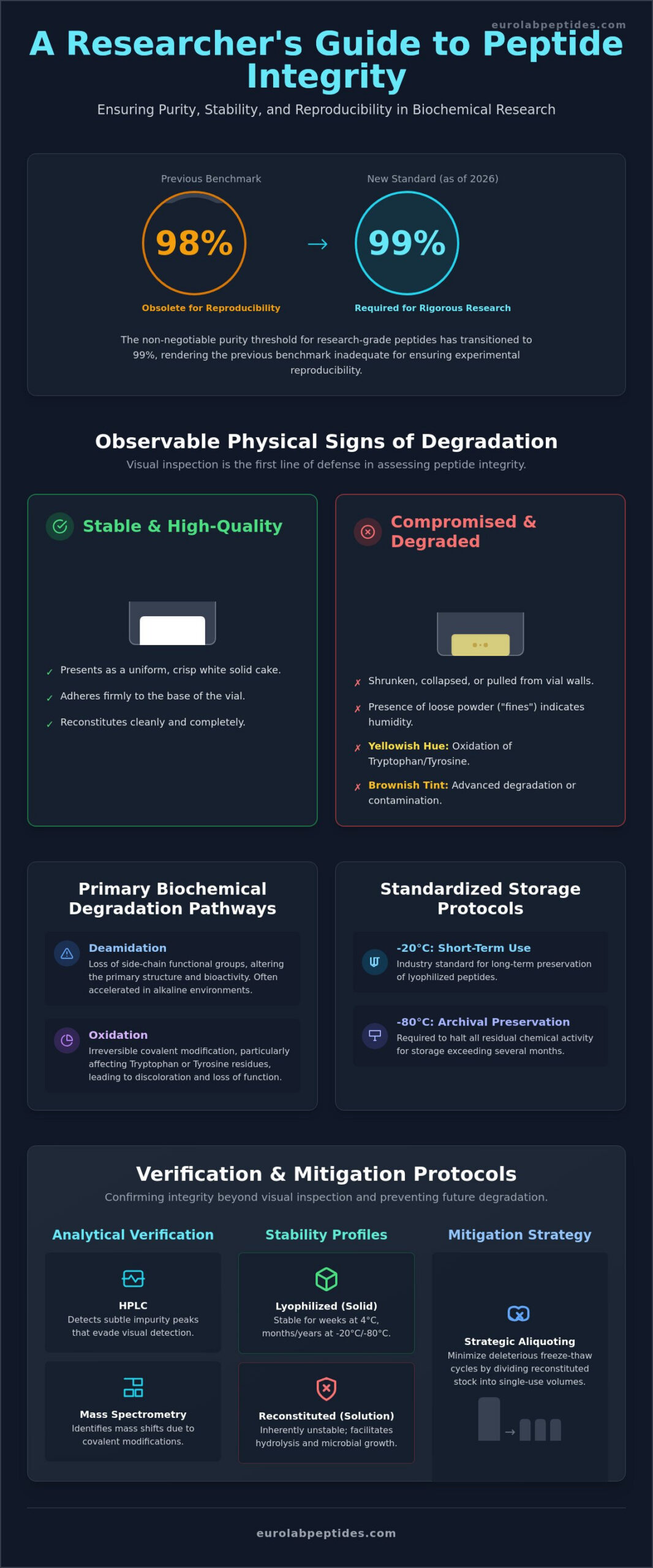
The non-negotiable purity threshold for research-grade peptides has transitioned to 99% as of 2026, rendering the previous 98% benchmark obsolete for ensuring rigorous experimental reproducibility. Maintaining peptide stability in research is no longer a passive assumption; it’s a variable that requires active, data-driven verification to prevent the introduction of bioactive impurities into in-vitro systems. You recognize that even minor deviations in the physical state of a lyophilized cake can signal a compromise in molecular integrity. The presence of compromised reagents frequently results in irreproducible data and the inefficient allocation of research budgets.
This clinical guide identifies the specific visual, chemical, and experimental markers of compromised peptide integrity to ensure laboratory accuracy. It provides a comprehensive checklist of visual red flags, examines primary chemical degradation pathways, and outlines a standardized protocol for verifying batch-specific purity. By adhering to these updated technical frameworks, researchers can maintain the high-stakes precision required for sophisticated biochemical inquiry.
Key Takeaways
- Identify macroscopic indicators of degradation, such as the transition from a uniform lyophilized cake to shrunken or discolored powder.
- Analyze how sequence-specific risks like deamidation and oxidation alter the primary structure and bioactivity of research-grade peptides.
- Maintain peptide stability in research by implementing standardized storage protocols at -20°C for short-term use and -80°C for archival preservation.
- Utilize High-Performance Liquid Chromatography and Mass Spectrometry to detect subtle mass shifts and impurity peaks that evade visual detection.
- Implement strategic aliquoting to minimize deleterious freeze-thaw cycles and ensure the long-term integrity of reconstituted reagents.
The Fundamentals of Peptide Stability and Integrity in Research
In the context of biochemical analysis, peptide stability in research is defined as the rigorous maintenance of a molecule’s primary sequence and its specific chiral integrity. Any deviation from the intended sequence, whether through the loss of side-chain functional groups or the cleavage of the backbone, constitutes a failure of stability. The peptide bond stability is the foundational metric here. The covalent linkage between the alpha-amino group of one amino acid and the alpha-carboxyl group of another remains vulnerable to nucleophilic attack, particularly in the presence of residual moisture. Maintaining this bond is critical for data accuracy.
Lyophilization serves as the primary defense against these kinetic processes. By removing water via sublimation, the peptide is transitioned into a low-energy, amorphous solid state. This process effectively minimizes the molecular mobility required for moisture-driven reactions. From a thermodynamic perspective, temperature fluctuations act as a catalyst for entropy. They provide the activation energy necessary for spontaneous bond cleavage. It’s essential to distinguish between physical degradation, such as non-covalent aggregation or precipitation, and chemical degradation, which involves irreversible covalent modifications like deamidation or oxidation.
Lyophilized vs. Solution Peptides: Stability Profiles
Aqueous environments are inherently unstable for long-term storage. They facilitate hydrolysis and provide a medium for microbial proliferation. The pH of the solution significantly dictates the rate of degradation. Alkaline environments often accelerate base-catalyzed deamidation. While lyophilized powders may remain stable for several weeks at 4°C, the industry standard for long-term research preservation is -20°C. For archival purposes exceeding several months, -80°C is required to halt all residual chemical activity. Solubility profiles often shift when stability is compromised. Solutions are high risk.
The Impact of Compromised Integrity on In-Vitro Models
Compromised reagents introduce significant noise into experimental data. When peptides undergo degradation, the resulting fragments or modified sequences often act as competitive inhibitors in cellular assays. This interference can lead to false-negative results or an artificial reduction in observed potency. Because the effective molar concentration of the active ligand is reduced, the reproducibility of the in-vitro model is fundamentally undermined. Strict handling protocols are mandatory. This ensures that the data generated reflects the intended biological interaction rather than an artifact of chemical decay.
Observable Physical Signs of Peptide Degradation
Visual inspection serves as the primary diagnostic tool for assessing the initial integrity of a sample. A stable, high-quality lyophilized cake should present as a uniform, crisp white solid that adheres to the base of the vial. When the cake appears shrunken, collapsed, or pulled away from the glass walls, it often indicates moisture ingress or exposure to temperatures exceeding the specific glass transition point. This physical collapse is rarely a benign change; it’s a precursor to the covalent decay that undermines peptide stability in research. Such structural failures suggest that the vacuum seal may be compromised, allowing atmospheric humidity to catalyze hydrolytic reactions.
Colorimetric shifts provide specific diagnostic data regarding the biochemical state of the reagent. While research-grade peptides are predominantly white, the emergence of yellowish hues is typically correlated with the oxidation of aromatic amino acids, specifically Tryptophan or Tyrosine residues. A transition toward a brownish tint indicates more advanced degradation or the presence of Maillard-like reactions with residual contaminants. These visible changes in the electromagnetic absorption of the sample are empirical evidence that the chemical and physical stability of the molecule has been breached. Researchers should also document the presence of “fines,” which are loose powder particles in a previously solid cake, as these suggest mechanical friability induced by humidity.
Changes in Reconstitution Behavior
The behavior of a peptide during reconstitution serves as a functional assay for its structural integrity. Reagents that previously achieved full dissolution within seconds but now require extended agitation or sonication are likely undergoing sub-visible structural transformations. Cloudiness or opalescence in a reconstituted solution suggests the formation of large-scale peptide aggregates. These aggregates are not merely inactive; they can introduce significant variability into in-vitro models by masking binding sites or triggering non-specific cellular responses. Utilizing high-purity reagents with verified stability profiles is essential to avoid these experimental artifacts.
A specific red flag is the “oil droplet” effect, where hydrophobic aggregates form, leading to incomplete solubility and visible droplets that refuse to homogenize. For instance, regenerative sequences such as bpc 157 must be monitored for these solubility shifts. Any deviation from a perfectly clear, colorless solution upon reconstitution indicates a loss of molar concentration. If particulates begin to form after an initially successful reconstitution, the peptide is precipitating out of the solution. This post-reconstitution precipitation fundamentally alters the intended dosage and renders the resulting data scientifically invalid.
Primary Biochemical Degradation Pathways: Sequence-Specific Risks
While visual assessments provide a preliminary baseline, the functional integrity of a sample is dictated by specific biochemical transformations. Peptide stability in research is frequently compromised through covalent modifications that are invisible to the naked eye but catastrophic to experimental validity. These pathways are sequence-dependent. The primary structure of the peptide dictates its inherent vulnerability to environmental stressors. Identifying these risks requires an understanding of how specific amino acid residues interact with their microenvironment over time.
Deamidation represents one of the most prevalent degradation pathways. It involves the hydrolytic conversion of Asparagine (Asn) and Glutamine (Gln) residues into Aspartate and Glutamate. This transformation introduces a negative charge and a slight mass increase, which can shift the isoelectric point and alter the peptide’s interaction with cellular targets. Similarly, hydrolysis results in the irreversible cleavage of the peptide backbone. Acid- or base-catalyzed cleavage often occurs at specific sites, such as the Aspartate-Proline (Asp-Pro) linkage, resulting in fragmented sequences that lack biological activity. Racemization, the loss of chiral integrity, is perhaps the most insidious pathway. It involves the conversion of L-amino acids to D-isomers, which preserves the molecular weight but fundamentally disrupts the spatial orientation required for receptor binding.
Oxidation of Cys and Met Residues
Methionine (Met) and Cysteine (Cys) residues are exceptionally vulnerable to oxidative stress. The mechanism typically involves the formation of methionine sulfoxide or the shuffling of disulfide bridges. Atmospheric oxygen and exposure to ambient light during laboratory handling are the primary environmental triggers for these reactions. Oxidation significantly alters the binding affinity in research peptides, as the addition of oxygen atoms changes the hydrophobicity and steric profile of the side chains. These modifications can transform a potent agonist into an inactive or inhibitory species, undermining the reliability of in-vitro models.
Deamidation and Iso-Aspartate Formation
The “Asn-Gly” sequence hazard represents a significant risk; the absence of a bulky side chain in Glycine facilitates the formation of a cyclic succinimide intermediate. This intermediate hydrolyzes to form a mixture of Aspartate and Iso-Aspartate, the latter of which introduces a “kink” into the peptide backbone. Maintaining a slightly acidic environment, typically between pH 5 and 6, is recognized as the optimal standard for stability. Detecting these subtle shifts in molecular weight and net charge requires advanced analytical methods, such as high-resolution mass spectrometry, to ensure the purity of the experimental reagent remains within the required 99% threshold.
Analytical Verification: Confirming Integrity Beyond Visual Inspection
Visual assessments are insufficient for confirming the 99% purity threshold required by modern laboratory standards. Analytical verification represents the final tier of quality control, providing the empirical data necessary to validate peptide stability in research. While a lyophilized cake may appear physically intact, sub-visible chemical transformations can only be resolved through high-resolution instrumentation. High-Performance Liquid Chromatography (HPLC) and Mass Spectrometry (MS) are the primary tools for identifying these deviations from the baseline established during initial synthesis.
Interpreting HPLC Chromatograms for Degradation
HPLC serves as the definitive method for quantifying purity by resolving the target molecule from its related substances. The emergence of “shoulder” peaks on a chromatogram is a diagnostic indicator of closely related impurities, such as deamidated species or diastereomers that elute near the parent peak. A broadening of the primary peak suggests conformational heterogeneity or the presence of soluble aggregates. Researchers must compare current elution profiles against the original Certificate of Analysis (CoA). Any shift in retention time or an increase in the area under the curve (AUC) for secondary peaks confirms that the chemical integrity has been breached. Access stability-tested reagents with batch-specific documentation to ensure analytical consistency in your protocols.
Mass Spectrometry provides the molecular weight confirmation required to identify the specific nature of the degradation. A mass shift of +16 Da is the classic signature of Methionine or Cysteine oxidation. Conversely, a +1 Da shift indicates deamidation, where the loss of an amine group and the addition of a hydroxyl group subtly alters the molecular mass. These shifts are often accompanied by unexplained changes in the background signal or baseline noise in sensitive assays. Such analytical red flags indicate that the effective concentration of the active peptide has been compromised.
Experimental Red Flags in Laboratory Data
Degradation often manifests as a loss of dose-response linearity in metabolic or longevity research models. When a peptide reagent is compromised, the actual molar concentration becomes an unknown variable, leading to an increased standard deviation across replicates. For example, ipamorelin studies require consistent purity to maintain valid GH secretagogue data; any impurity-driven interference can produce divergent results that undermine the entire data set. If the same protocol yields inconsistent results across different batches of the same stock, the investigator should immediately suspect a failure in peptide stability in research. Maintaining high-stakes precision requires the rejection of any reagent that fails to meet these analytical benchmarks.
Mitigating Degradation: Protocols for Sourcing and Storage
Preserving peptide stability in research requires a transition from passive storage to active environmental management. Standardized protocols mandate that lyophilized powders are maintained at -20°C for short-term applications. For archival storage exceeding four weeks, -80°C is the non-negotiable requirement to halt residual chemical activity. Exposure to atmospheric moisture and ambient light must be strictly controlled through the use of amber vials and desiccated storage environments. These measures prevent the initiation of the oxidative and hydrolytic pathways that compromise molecular integrity. Maintaining these specific conditions is the only way to ensure that the primary sequence remains unchanged from the point of synthesis.
Procedural handling during the initial laboratory entry is equally critical. An effective aliquoting strategy involves dividing reconstituted solutions into single-use volumes immediately after dissolution. This approach minimizes the number of freeze-thaw cycles, each of which introduces thermodynamic stress that facilitates aggregation and bond cleavage. By isolating the bulk stock from repeated thermal fluctuations, researchers maintain the effective molar concentration of the reagent throughout the duration of the study. It’s a fundamental step that protects the investment in high-purity reagents and ensures that experimental data remains reproducible across different time points.
The EuroLab Multi-Level Quality Protocol
The selection of a reliable source is the first step in ensuring peptide stability in research. EuroLab Peptides utilizes a multi-level quality control process that includes both in-house analysis and independent third-party laboratory verification. This rigorous oversight ensures that every batch meets the 99% purity threshold before it enters the supply chain. Regional European logistics are optimized to meet the temperature-controlled shipping standards for 2026, reducing the risks associated with prolonged transit and international customs delays. Every shipment is accompanied by batch-specific documentation, providing researchers with the empirical validation required for high-stakes precision inquiry. This transparency allows for the immediate integration of reagents into validated experimental frameworks without the uncertainty of unverified purity.
Best Practices for Handling and Reconstitution
The reconstitution phase represents a high-risk window for chemical decay. Utilizing sterile, degassed buffers is a mandatory requirement to eliminate dissolved oxygen, which serves as a primary catalyst for the oxidation of Methionine and Cysteine residues. Researchers must prioritize gentle swirling over vigorous vortexing during the homogenization process. Vortexing generates mechanical shearing forces and introduces air bubbles, both of which can lead to the denaturation of complex sequences or the formation of insoluble aggregates. To ensure the accuracy of your data, it’s essential to secure high-purity research peptides backed by verifiable analytical data and regional logistics support. Adhering to these clinical handling standards ensures that the final experimental results remain a true reflection of biological activity rather than a consequence of reagent degradation.
Advancing Experimental Precision Through Rigorous Stability Standards
EuroLab Peptides provides this security through a multi-level quality control process and independent third-party laboratory verification. Every batch is supported by specialized European-based logistics to maintain temperature stability during transit. You can Order Third-Party Tested Research Peptides from EuroLab to secure the integrity of your next study. Implementing these clinical benchmarks ensures that your data reflects biological reality rather than reagent artifacts. Your commitment to these rigorous standards will yield more reproducible and impactful results.
Frequently Asked Questions
Can I still use a peptide if the solution is slightly cloudy?
Reconstitution resulting in a cloudy or opalescent solution indicates large-scale peptide aggregation or microbial proliferation. Such physical changes signify that the molecular integrity is compromised and the effective molar concentration is no longer accurate. Utilizing cloudy solutions introduces significant noise into in-vitro models and can lead to non-specific cellular responses. Reagents showing any degree of turbidity should be discarded to maintain experimental rigor.
How much does one freeze-thaw cycle impact peptide stability?
A single freeze-thaw cycle introduces significant thermodynamic stress that can initiate both physical and chemical degradation. These thermal fluctuations facilitate the formation of insoluble aggregates and can accelerate the rate of covalent bond cleavage in aqueous environments. Dividing reconstituted peptides into single-use aliquots before the initial freeze is the only reliable method to prevent the cumulative damage caused by repeated transitions between solid and liquid states.
What is the most common chemical sign of peptide degradation in storage?
Deamidation of Asparagine and Glutamine residues represents the most frequent chemical transformation during storage. This hydrolytic process results in the formation of Aspartate or Iso-aspartate, which alters the peptide’s net charge and spatial orientation. Oxidation of Methionine and Cysteine residues is also prevalent, particularly when samples are exposed to atmospheric oxygen or ambient light. These modifications are primary drivers of reduced peptide stability in research.
Does the color of the lyophilized powder always indicate its purity?
Color serves only as a macroscopic indicator and is not a definitive metric for purity. While a yellowish tint often suggests the oxidation of Tryptophan or Tyrosine, many covalent modifications occur without altering the visual appearance of the white lyophilized cake. Deamidation and racemization frequently leave the physical state of the powder unchanged. Analytical verification via HPLC remains the only method to confirm that a sample meets the 99% purity threshold.
How long do research peptides typically last once reconstituted?
Reconstituted peptides are inherently less stable than their lyophilized counterparts and typically maintain integrity for only several days at 4°C. Aqueous environments facilitate rapid hydrolysis and provide a medium for microbial growth. If the solution is not utilized within 24 hours, it must be aliquoted and stored at -20°C or -80°C to minimize decay. Stability profiles vary significantly based on the specific amino acid sequence and the pH of the buffer used.
Which amino acid sequences are most prone to rapid degradation?
Sequences containing the Asparagine-Glycine (Asn-Gly) motif are exceptionally vulnerable to rapid deamidation via a cyclic succinimide intermediate. Additionally, the Aspartate-Proline (Asp-Pro) linkage is highly susceptible to acid-catalyzed backbone cleavage. Peptides rich in Methionine, Cysteine, or Tryptophan are at high risk for oxidative damage. Understanding these sequence-specific risks is essential for researchers handling complex synthetic molecules in longitudinal studies.
Can mass spectrometry detect all forms of peptide degradation?
Mass spectrometry is highly effective for detecting mass shifts associated with oxidation (+16 Da) or deamidation (+1 Da), but it cannot detect racemization. Racemization involves the conversion of L-amino acids to D-isomers, which results in no change to the molecular weight. Identifying these chiral shifts requires High-Performance Liquid Chromatography (HPLC) or specialized chiral analysis. Relying solely on mass data can lead to a false sense of security regarding total molecular integrity.
Is it possible for a peptide to degrade without any visual changes?
Degradation often occurs at the molecular level without any observable change to the lyophilized cake or the clarity of the reconstituted solution. Subtle covalent modifications can fundamentally alter binding affinity and biological activity while the reagent remains a crisp white powder. This underscores why peptide stability in research cannot be assessed by visual inspection alone. Independent third-party validation and batch-specific HPLC data are required to ensure the reagent remains viable for precise inquiry.
