

A detectable signal isn’t, by itself, evidence of meaningful binding. In vitro peptide binding assays test whether a peptide interacts with a target under defined conditions, but the result depends on the research question, assay format, and experimental setup. Labelling or immobilizing a molecule may change how it behaves, while nonspecific binding can produce a signal that is difficult to interpret.
Choosing a method is more than a technical preference. First decide what evidence you need: binding kinetics, a comparison of relative interactions, or a screen for candidate peptides. Appropriate controls and consistent assay conditions help distinguish a reproducible interaction from an artifact. No single readout establishes biological significance on its own.
This article compares common approaches, including surface plasmon resonance (SPR) and microscale thermophoresis (MST), and explains where each can be useful. You’ll learn how to match a method to your research question, identify potential sources of bias, and plan controls that support careful interpretation.
Key Takeaways
- Use in vitro peptide binding assays to investigate direct peptide-target interactions, and distinguish those results from functional readouts or computational predictions.
- Compare SPR, biolayer interferometry, fluorescence polarization, and isothermal titration calorimetry by their measurement principles and practical limitations.
- Match the assay to your research question, target format, peptide properties, and available instrumentation.
- Use scientifically appropriate positive, negative, and reference controls to identify nonspecific signals and interpret results cautiously.
- Consider material identity, purity, and consistency as part of a reproducible workflow, while keeping conclusions specific to the evidence measured.
What Do In Vitro Peptide Binding Assays Measure?
An in vitro peptide binding assay examines whether a peptide interacts with a molecular target outside a living organism. The experiment brings the molecules together in a defined system and measures a change associated with their interaction. Depending on the method, the readout may reflect association or dissociation, or a change in fluorescence or refractive index. The result applies to the conditions tested, including target format, buffer, temperature, and concentrations.
Binding is an interaction detected under defined experimental conditions. Affinity describes the strength of that interaction as estimated by a method-specific measurement. Functional activity is a biological effect following an interaction and must be assessed separately.
This distinction matters because direct binding data and downstream functional readouts answer different questions. A cell-based response may indicate a biological effect without proving direct peptide-target contact. Conversely, a measured interaction may not alter target function. Computational predictions can help prioritize hypotheses, but they are not experimental confirmation. The broader principles of ligand-binding measurements are summarized in this Ligand Binding Assay overview.
Binding, affinity, and selectivity are different questions
Binding asks whether an interaction is observed in the chosen assay. Affinity asks how strongly the molecules interact, often reported as a method-dependent estimate such as an equilibrium dissociation constant. Selectivity asks whether a peptide binds one target more strongly or consistently than related targets, such as a receptor family member or structurally similar protein. Evidence of binding alone does not establish high affinity or selectivity.
Where peptide binding assays fit in research
Researchers use binding measurements to test target-interaction hypotheses, compare peptide variants, or examine how changes to a target affect interaction. Peptide libraries provide collections of candidates for screening; they describe what is being tested, not a separate measurement principle. Foundational terminology about peptide structure and composition can help frame these experiments, as outlined in this guide to what peptides are.
A positive assay signal is evidence only within the limits of the design. It does not, by itself, establish biological relevance, target selectivity, or activity in vivo. For example, a peptide may associate with an immobilized target in vitro yet fail to engage the same target in a more complex system. Nonspecific interactions, surface effects, aggregation, and detection artifacts can also contribute to a signal. Interpret results in light of controls, reproducibility, and whether the measured interaction addresses the research question.
Before treating a result as evidence, identify what the assay directly measures:
- Direct interaction: Does the measurement support physical association between the peptide and target?
- Comparative behavior: Were related targets or peptide variants assessed under comparable conditions?
- Functional consequence: Was a separate experiment used to test whether binding changes target activity?
Keeping these conclusions separate makes in vitro peptide binding assays more informative. The data can support a defined interaction claim without overstating what they establish about biological function.
How Common In Vitro Peptide Binding Assays Generate a Signal
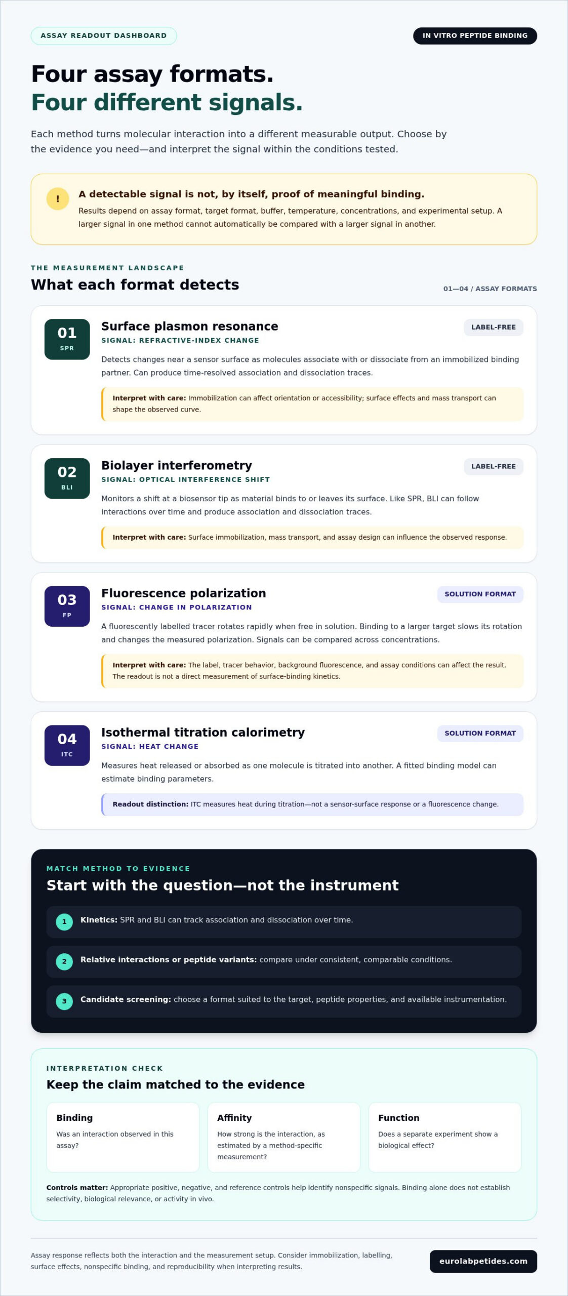
Each assay converts a molecular interaction into a measurable output, but the output depends on the method’s physical principle. Some formats track changes at a sensor surface; others measure altered fluorescence or heat in solution. Because the readouts differ, a larger signal in one method cannot automatically be compared with a larger signal in another.
Label-free methods: SPR and biolayer interferometry
Surface plasmon resonance (SPR) detects changes in refractive index near a sensor surface as molecules associate with or dissociate from an immobilized binding partner. Biolayer interferometry (BLI) monitors shifts in an optical interference pattern at a biosensor tip as material binds to or leaves its surface. Both can follow interactions over time, producing association and dissociation traces rather than only a final endpoint.
These time-resolved data can support kinetic estimates, but interpretation depends on assay design and model fit. Immobilization may affect molecular orientation or accessibility, while surface effects and mass transport can shape the observed curve. The signal may reflect the delivery of molecules to the surface as well as the interaction itself, so sensor response is not an unqualified measure of binding strength.
Solution and signal-based formats
Fluorescence polarization (FP) commonly tracks a fluorescently labelled small molecule or peptide. A free tracer rotates rapidly in solution; when it binds to a larger target, its rotation slows and the measured polarization changes. FP can be used to compare signals across concentrations, but the label, tracer behavior, background fluorescence, and assay conditions can affect the result. The readout reflects a change in rotation, not a direct measurement of surface-binding kinetics.
Isothermal titration calorimetry (ITC) measures heat released or absorbed as one molecule is titrated into another. A fitted binding model can estimate parameters such as affinity, stoichiometry, and enthalpy. Because ITC detects heat rather than a label, it avoids fluorophore modification. However, the interaction must produce a resolvable heat change under the chosen conditions, and dilution heat and sample properties also need consideration.
Method-specific readouts require method-specific interpretation: each signal reflects a distinct physical measurement, not a universal unit of binding. Before selecting among these in vitro peptide binding assays, decide whether you need time-resolved kinetics, a concentration-dependent fluorescence response, or thermodynamic characterization. Then consider target stability, peptide solubility, tolerance for labelling, sample requirements, and available instrumentation. Every format still requires assessment of background and assay-specific artifacts.
Reproducible measurements also depend on consistent research materials and clear documentation. EuroLab Peptides uses in-house and independent third-party laboratory testing as part of its quality-control process for research-grade peptides. Its products are intended strictly for in vitro research and laboratory development. When planning an experiment, consider research-grade peptides for laboratory research alongside the assay’s material and documentation requirements.
How to Compare Assays for Peptide-Target Binding
Select a method based on the evidence you need, not simply on which instrument is available. Affinity estimation, kinetic characterization, and primary screening are different aims. A platform suited to following association and dissociation may not be practical for screening a large peptide set. A measurable response also does not make unlike assay outputs directly comparable.
| Method | Measurement principle and sample format | Strengths | Interpretive caveats |
|---|---|---|---|
| SPR | Tracks refractive-index changes near a sensor surface, typically with one binding partner immobilized. | Time-resolved traces can support association and dissociation analysis. | Immobilization, surface behavior, and mass transport can influence the observed response. |
| BLI | Measures optical interference shifts at a biosensor as molecules bind or dissociate; commonly uses a surface-bound partner. | Provides time-course measurements and can compare interaction profiles. | Sensor loading, surface effects, and sample behavior may affect fitted results. |
| Fluorescence polarization | Measures changes in rotation of a fluorescent tracer in solution as it binds a larger molecule. | Can support concentration-dependent comparisons in a solution format. | Tracer labelling, fluorescence background, and peptide properties can alter signal quality. |
| ITC | Measures heat changes during titration of one binding partner into another in solution. | Can provide thermodynamic parameters, including binding enthalpy and stoichiometry, under suitable conditions. | Requires a resolvable heat signal; dilution effects and sample requirements matter. |
Match the method to the interaction question
For kinetic characterization, SPR and BLI monitor association and dissociation over time, although results remain sensitive to surface-based assay design. For thermodynamic characterization, ITC measures heat and can provide energetic information that a sensor response alone does not. Primary screening instead prioritizes throughput and practical sample use. Treat screening hits as candidates for follow-up confirmation, not as conclusive evidence based on signal intensity alone.
Account for peptide and target-format constraints
Before choosing a format, consider peptide size, solubility, aggregation tendency, and whether a fluorescent label could alter binding. Assess the target too: immobilization or modification may mask an interaction site or change its accessibility. A soluble target may suit one approach, while a stable, accessible surface-bound target may suit another. Material identity, purity, and consistency also support interpretable comparisons; see the guide to third-party tested peptides.
Instrument access, sample quantity, and the need for kinetic, thermodynamic, or screening data all affect feasibility. No platform is universally optimal. Choose in vitro peptide binding assays to fit the target format and study design, and plan follow-up experiments that address the specific claim.
How to Design and Interpret a Peptide Binding Experiment
A defensible experiment begins with a precise claim. Decide whether you’re testing for a detectable interaction, estimating affinity, comparing selectivity, or screening candidates. Then choose an assay whose readout addresses that question rather than treating every signal as equivalent evidence. A practical workflow is:
- Define the question. State which peptide-target interaction or comparison the experiment will examine.
- Select the format. Match the method to the target’s presentation, peptide properties, and intended measurement.
- Validate materials. Document identity, concentration, storage, preparation, and relevant target properties.
- Run controls. Include controls suited to evaluating assay responsiveness, nonspecific signal, and target dependence.
- Interpret within scope. Report what the data demonstrate, and separate that observation from broader mechanistic claims.
In in vitro peptide binding assays, a control is useful when it tests a defined source of uncertainty. A positive control can show that the assay responds to a known interaction under the chosen conditions. A negative control, such as a suitable nonbinding peptide or target-free condition, can help estimate background or nonspecific signal. A reference control may help compare runs or conditions. Choose controls for the assay and biological system; no single design fits every experiment.
Controls and validation that strengthen interpretation
Evaluate background before interpreting the test sample. Apply background correction consistently and retain raw measurements so the correction is transparent. Replicates help establish whether a pattern is reproducible, not simply provide more measurements to average. If a replicate is excluded, report why and which criterion was used. Where practical, use an orthogonal method with a different measurement principle to test whether the finding depends on one assay format.
A binding claim should be supported by appropriate controls and, where feasible, orthogonal evidence. Report the conditions needed to interpret the result, including concentrations, buffer, temperature, incubation conditions, target format, detection settings, replicate structure, and analysis approach. State limitations directly. This record helps readers distinguish observations from conclusions that depend on assumptions.
Common sources of ambiguity in binding data
Nonspecific adsorption to assay surfaces can resemble target-dependent binding. Peptide aggregation may alter apparent concentration or produce irregular signals, while assay interference can affect detection without reflecting the interaction of interest. These possibilities matter when a signal changes with concentration but controls do not establish the reason. Examine background behavior and replicate consistency alongside the main response.
Signal magnitude alone cannot establish specificity or biological function. A strong response may reflect assay geometry, target presentation, or interference; a modest response may still be meaningful within a validated format. Keep conclusions in three layers: what was observed, what mechanism the data support, and what remains an extrapolation to other experimental systems. Research-grade materials intended strictly for in vitro research and laboratory development can support controlled studies, but sourcing does not replace experimental validation.
For research-grade peptides intended for laboratory research, explore research-grade peptide options.
From Assay Selection to Research-Grade Peptide Sourcing
A defensible workflow connects the experimental question to the assay, its controls, and the materials being tested. Method limitations shape what can be concluded; material identity and consistency help determine whether differences between runs reflect the experiment or variation in the input. These elements work together. An appropriate platform cannot compensate for poorly characterized materials, and material documentation cannot establish binding on its own.
Identity, purity, and consistency matter because impurities or batch variation may affect sample concentration, background, or reproducibility. They are quality considerations, not evidence that a peptide will interact with a particular target. A purity result alone does not demonstrate binding activity, selectivity, or biological function.
Document materials and methods for reproducibility
For each experiment, record peptide identity and batch information, relevant material documentation, storage history, preparation details, and assay conditions. Include target format, concentrations, buffer, temperature, and analysis approach in the experimental record. This context makes results easier to compare across runs and helps identify variables that may explain a discrepancy. Quality-control documentation supports traceability; it does not guarantee experimental performance.
Research-material sourcing is one part of assay planning. EuroLab Peptides uses in-house and independent third-party laboratory testing as part of its quality-control process. This information can inform material evaluation alongside the requirements of the chosen assay and relevant research documentation. It does not prove that a specific peptide is suitable for a particular target or method.
Continue with an evidence-led research workflow
Before beginning, compare the assay’s sample and detection requirements with the research-material information for the peptide. Consider whether the method requires immobilization, labelling, or particular sample properties, then document how the material was handled and used. These practices help make in vitro peptide binding assays more reproducible and their conclusions easier to assess, while keeping claims within the evidence generated.
Use research-grade peptides strictly for in vitro research and laboratory development. A measured interaction applies to the tested conditions; it does not establish clinical applicability or predict performance in a living system. Clear records of materials and methods make that boundary explicit and preserve the distinction between an assay result and a broader biological claim.
For research-grade materials intended for laboratory development, explore EuroLab Peptides.
Build Your Next Assay on a Defensible Foundation
Turn the experimental question into a clear plan before committing samples: define the claim, identify the evidence needed to support it, and decide what documentation will make the work reproducible. A well-designed study treats material selection as one part of the plan, alongside assay conditions, controls, and transparent interpretation.
Traceability can help researchers assess consistency across experiments. EuroLab Peptides uses in-house and independent third-party laboratory testing as part of its quality-control process, and its products are designated strictly for in vitro research and laboratory development. These practices inform material evaluation; they do not establish binding or predict assay outcomes. Keep conclusions anchored to data from the chosen method.
As you plan future in vitro peptide binding assays, prioritize a workflow in which each measurement answers a defined question and each claim is supported by appropriate evidence. Explore EuroLab Peptides research materials for laboratory research. A careful plan is a practical starting point for more interpretable research.
Frequently Asked Questions
What is an in vitro peptide binding assay?
An in vitro peptide binding assay is a laboratory test used to evaluate peptide interaction with a target in a controlled, nonliving system. For example, a researcher might test whether a candidate peptide associates with a purified protein under specified buffer and concentration conditions. The result applies to that setup. It does not, by itself, show that the interaction occurs in cells, changes target function, or has an effect in an organism.
Which assay is best for measuring peptide binding?
No single method is best for every peptide-target pair. A study focused on association and dissociation rates may suit a time-resolved surface method, while fluorescence polarization may be useful if a fluorescent tracer can be used without disrupting the interaction. Consider target format, peptide solubility, sample availability, and instrumentation. Choose the method whose output answers the research question, then plan how to validate the finding.
Can fluorescence polarization measure peptide binding?
Yes. Fluorescence polarization can detect binding by measuring how the rotation of a fluorescent tracer changes as it associates with a larger molecule. A researcher can test a range of target concentrations and assess whether the signal changes consistently. The tracer’s label and behavior matter: a label may affect the interaction, and fluorescence interference can complicate measurement. Include appropriate controls and interpret the result as a polarization change, not as direct kinetic data.
How do researchers confirm a peptide-target interaction?
Researchers can strengthen evidence for a proposed interaction by testing a concentration series, checking that the response depends on the target, and reproducing the pattern in independent runs. A competition experiment, in which an unlabeled candidate is tested against a labelled binding partner, may provide supporting evidence if the assay supports it. Confirmation with a second method based on a different measurement principle can help reveal format-specific artifacts. Keep conclusions limited to the evidence collected.
What controls are important in peptide binding assays?
Useful controls depend on the assay, but often include a positive control to verify that the system can detect a known interaction and a negative control to assess background or nonspecific signal. A target-free condition can help identify peptide interaction with the assay surface or detection system. Reference samples may help compare runs. Define control purposes in advance, apply background correction consistently, and report unexpected control behavior rather than excluding it without explanation.
Can peptide library screening prove that a peptide binds a target?
No. Library screening identifies candidates that produce a signal under screening conditions; it does not establish that each candidate binds specifically or reproducibly. For example, a sequence selected from a screen may need to be synthesized or isolated individually, tested across a concentration range, and evaluated against suitable negative or related-target controls. Follow-up with an independent assay can further assess whether the signal reflects the proposed interaction rather than a screening-format artifact.
