

The key difference is structural, not just a change in name: IGF-1 LR3 contains 83 amino acid residues, including a 13-residue N-terminal extension and an arginine substitution at position 3, while native human IGF-1 contains 70 amino acids. For IGF-1 LR3 for laboratory research, those changes matter because they affect how the molecule interacts with receptors and IGF-binding proteins.
That distinction can be easy to overlook when comparing related analogs, yet it shapes how researchers interpret binding, bioavailability, and experimental results. This article explains how IGF-1 LR3 differs from native IGF-1 and IGF-1 DES, what reduced binding to IGF-binding proteins means, and how analytical testing can assess identity, purity, and quantity. It also clarifies why potency and binding characteristics should be considered when designing and interpreting laboratory protocols.
Key Takeaways
- Compare the molecule’s sequence changes with native IGF-1 to understand why it behaves differently in experimental systems.
- Distinguish IGF-1 LR3 from IGF-1 DES by their structural differences, not just their names.
- Separate IGF-1 receptor binding from insulin receptor binding when interpreting signaling data.
- Account for IGF-binding proteins when assessing how much IGF-1 may remain bioavailable in a given research context.
- Use analytical evidence for identity, purity, and quantity to evaluate whether a sample is suitable for a study.
Precise difference between IGF-1 LR3 and native IGF-1?
IGF-1 LR3 differs from native IGF-1 through an N-terminal extension and an amino-acid substitution. These structural changes affect how the analog interacts with binding proteins and matter when interpreting experimental activity.
Amino-Acid Modifications in LR3
IGF-1 LR3 contains 83 amino acid residues, including a 13-residue extension at its N-terminus. It also has arginine at position 3, where native IGF-1 has glutamic acid. Together, these modifications distinguish the LR3 analog and explain its name.
For experimental comparisons, treat sequence identity as a key factor: the modified molecule is not interchangeable with native IGF-1. The U.S. regulatory distinction also matters: IGF-1 LR3 is not FDA-approved for human or veterinary use, and the FDA has issued a warning letter addressing IGF1-LR3 compounding concerns. These facts do not establish a comprehensive rule for every research purchase or possession.
Differences from Endogenous IGF-1
Native human IGF-1 is 70 amino acids long, while the described IGF-1 LR3 analog has 83 residues. Its extended sequence and substitution at position 3 make the molecular structures distinct, even though both are IGF-1 forms.
That difference is important when selecting an appropriate comparator or interpreting an assay. A response to LR3 cannot automatically be attributed to native IGF-1, because sequence changes can influence binding and biological action. Molecular mass can serve as a measurable identifier in analytical comparison.
IGF-1 Structure and Biological Action
IGF-1 is a peptide growth factor whose biological action depends on interactions with receptors and binding proteins. Structure provides context for those interactions, but it does not by itself predict the size or direction of an experimental response. This distinction matters in studies involving muscle cells or other model systems.
Research has examined IGF-I variants in pigs and marmoset monkeys, long R3 IGF-I infusion in guinea pigs, and IGF-1 LR3 with IGFBP-3 in L6 myogenic cells. A 2023 fetal-sheep study reported that attenuated glucose-stimulated insulin secretion during acute infusion did not persist in isolated islets. A 2025 study reported no growth promotion in late-gestation growth-restricted fetal sheep.
These findings describe specific experimental models, not a general therapeutic effect. They also do not establish a human exposure-toxicity profile. Potential hazards depend on exposure route, concentration, and biological context; receptor activity alone cannot define a safe threshold. IGF-1 LR3 is intended strictly for laboratory research, not human consumption, therapeutic use, diagnostic use, or veterinary use.
IGF-1 LR3 comparison with IGF-1 DES and native IGF-1
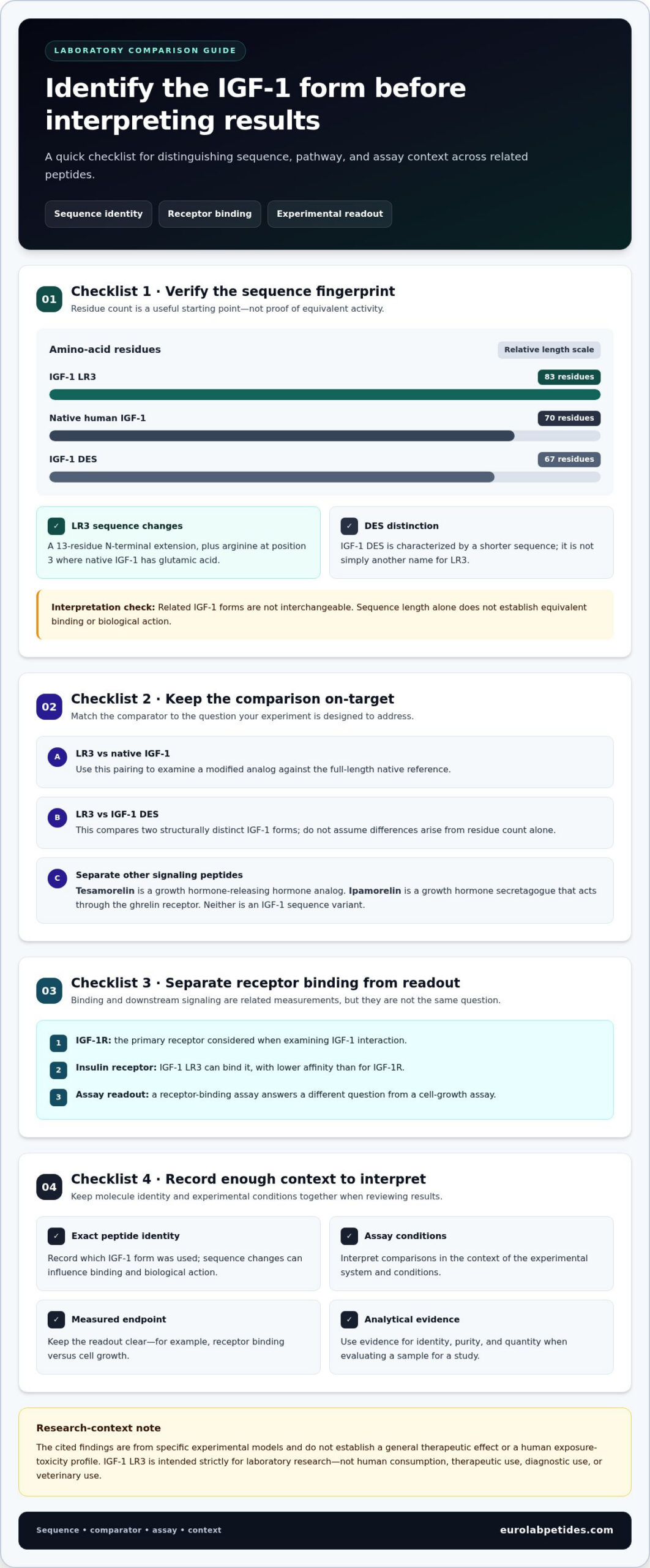
Structural length distinguishes these three IGF-1 forms: native human IGF-1 has 70 amino acids, IGF-1 DES has 67, and the described IGF-1 LR3 analog has 83. These are related peptides, but they are not interchangeable experimental reagents. Their sequence differences can affect molecular interactions, so a biological result from one form should not be assigned automatically to another. Comparison begins by identifying the exact molecule used.
For a scientific comparison, match the experimental system and readout before interpreting differences. A receptor-binding assay, for example, answers a different question from a cell-growth assay. Record peptide identity, assay conditions, and measured endpoint together; otherwise, apparent differences may reflect study design rather than an intrinsic distinction between analogs. Choose a comparator that fits the hypothesis, such as a study of sequence changes or receptor-related activity.
Native IGF-1 and IGF-1 DES
Native IGF-1 provides the full-length reference, while IGF-1 DES is a shortened form. The numerical sequence contrast is useful for sample identification, but length alone does not establish equivalent binding or biological action. A study comparing IGF-1 LR3 with native IGF-1 tests a different relationship than one comparing LR3 with DES. Keep those pairings separate when organizing controls or interpreting results.
IGF-1 DES and IGF-1 LR3 also differ in modification pattern, not simply in residue count. LR3 has an N-terminal extension and an arginine substitution at position 3; DES is characterized by a shorter sequence. Therefore, an observed response cannot be explained by length alone without examining the molecular structure and assay context. This distinction matters when researchers compare synthetic analogs with native material.
Tesamorelin and Ipamorelin
Tesamorelin and Ipamorelin are not IGF-1 sequence variants. Tesamorelin is a growth hormone-releasing hormone analog, while Ipamorelin is a growth hormone secretagogue that acts through the ghrelin receptor. Their biological action involves upstream hormonal signaling, unlike direct comparison among IGF-1 forms. Similar study themes do not make these peptides equivalent research materials.
Accordingly, an experiment comparing IGF-1 LR3 with Ipamorelin asks a different question from an LR3-versus-native IGF-1 comparison. The former contrasts distinct signaling pathways; the latter examines related IGF-1 molecules. Tesamorelin belongs in the same pathway-level distinction, not as a structural substitute. Separating these categories helps researchers interpret data without conflating molecular identity, receptor action, and regulation of upstream signals.
Does IGF-1 LR3 bind the insulin receptor?
Yes. IGF-1 LR3 can bind the insulin receptor, but its affinity is lower than for IGF-1R. The distinction matters because receptor binding and downstream signaling are related, but not identical, experimental measurements.
IGF-1 Receptor Interaction
IGF-1R is the primary receptor considered when examining IGF-1 interaction. IGF-1 LR3 maintains reported IGF-1R affinity at 0.5-1 nM. This value helps describe binding, but does not alone establish the strength of a biological response in a particular cell model.
For comparison, the stated affinity range for the IGF-1 receptor is approximately 10−8-10−9 M. Differences in assay conditions and receptor presentation affect how binding results should be interpreted. Researchers should therefore compare measurements produced with the same assay design, rather than treating values from different experimental systems as directly equivalent.
Binding is one part of receptor action. After ligand interaction, IGF-1R activation can initiate intracellular signaling, with the measured outcome depending on the cell type and assay endpoint. A binding assay can establish interaction; a separate functional assay is needed to assess signaling. Keeping those steps distinct prevents receptor affinity from being mistaken for a direct measure of cellular effect.
IGF and Insulin Receptor Signaling
IGF-1 LR3 also binds the insulin receptor, with 10 to 100 fold lower affinity than its affinity for IGF-1R. This comparison indicates a preference for IGF-1R, not an absence of insulin receptor interaction. The extended analog should therefore be assessed against both receptor targets when receptor selectivity is central to an experiment.
IGF-1R and the insulin receptor are related receptor systems, but their interaction with a ligand does not guarantee identical signaling. Receptor abundance, cell context, and the selected readout all shape an experimental result. For example, a binding result at IGF-1R cannot by itself establish the size of an insulin-receptor-mediated response.
A clear analysis separates three steps: identify the receptor, measure ligand binding, then assess downstream signaling. Applying this sequence to IGF-1 LR3 helps distinguish receptor affinity from functional activity. It also supports fair comparisons with native IGF-1, provided each ligand is tested under matching conditions and the specific receptor position is documented.
For study interpretation, report IGF-1R and insulin receptor results separately. A combined label such as “IGF receptor activity” can obscure the lower insulin-receptor affinity reported for LR3. This distinction is especially relevant when evaluating receptor selectivity or comparing an extended analog with another ligand in a defined cellular model.
IGF-binding proteins and IGF bioavailability
IGF-binding proteins (IGFBPs) regulate how IGF-1 is carried and presented to tissues. The six IGFBPs bind approximately 98% of circulating IGF-1, so total concentration and freely available peptide are not equivalent measures. For an experimental readout, that distinction affects how researchers interpret growth factor exposure and cellular activity.
Affinity helps explain this system. IGFBPs bind circulating IGF-1 with an affinity of approximately 10−10 M. IGFBP-3 accounts for around 80% of IGF-1 bound to IGFBPs. With IGF-1 and the acid-labile subunit, IGFBP-3 typically forms a 150 kDa complex. This assembly changes transport and persistence, rather than simply representing another receptor interaction.
IGF-1 LR3 has substantially reduced affinity for IGFBPs compared with native IGF-1. Its IGFBP affinity is described as reduced by 100-1000 fold, a structural consequence relevant to interpreting availability in a model. Reduced binding can alter the balance between protein-bound and unbound material. It does not, by itself, quantify receptor activation or predict the outcome of a particular assay.
Reported persistence values depend on the context measured. Natural IGF-1 is described as having a half-life of less than 20 minutes, while IGF-1 LR3 is reported to remain active in the bloodstream for 20 to 30 hours. A separate measurement reports a typical murine plasma half-life of 4-8 hours after subcutaneous injection. These values describe different endpoints or experimental settings and should not be treated as interchangeable.
That distinction matters when evaluating experimental exposure. A plasma half-life describes the decline of measurable material in a specified model; “active in the bloodstream” describes a broader activity claim. Neither directly establishes the duration of cellular signaling. For example, an in vitro assay with IGFBP-3 may produce different free-peptide availability than a system without that binding partner.
IGFBP-related proteins also bind IGFs, but with much lower affinity than the six IGFBPs. Distinguish their contribution from the high-affinity binding system. To interpret results clearly, identify the model, binding partners, and measured endpoint, then distinguish total peptide from bioavailable peptide. These factors help explain an observed response without treating reduced binding as proof of a specific clinical effect.
Purity, identity, quantity, and laboratory testing
For IGF-1 LR3 for laboratory research, evaluate identity, purity, quantity, and storage as separate quality attributes. Each addresses a different source of experimental variation, so one result cannot substitute for the others.
Purity, Identity, and Quantity Testing
Reverse-phase HPLC purity of at least 98% is the stated research-grade benchmark. HPLC estimates the proportion of target peptide relative to detected impurities, but it does not independently establish sequence identity or the total amount in a vial. Mass spectrometry provides identity evidence, while quantity requires its own assessment.
A lot-specific certificate of analysis should include HPLC purity, mass-spectrometry identity, and endotoxin results from an independent laboratory. The listed CAS number is 946870-92-4, which can be compared with the documentation. The recommended endotoxin level is below 1 EU/mg. These results address distinct questions, so read each result separately rather than treating a purity percentage as complete verification.
For studies requiring consistent material, match the lot identifier on the vial to the certificate and retain both with study records. A 5 mg vial is one stated example of lyophilized material, while a separate product listing gives a 1 mg strength. Those are different specifications, not interchangeable amounts. A multi-month study can reduce batch-to-batch variability by using material from one lot.
Analytical evidence also supports interpretation of structural and biological comparisons. If a sample’s identity or purity differs from its documentation, measured effects may reflect sample variation rather than the intended experimental factor. In IGF-1 LR3 for laboratory research, therefore, compare the certificate, label, and lot information before assigning results to a particular pathway or application.
Concentrations, Storage, and Stability
Reported in vitro concentrations range from 1 to 100 nM, with a typical working concentration of 10-100 ng/mL. After reconstitution, the recommended working stock range is 100 µg/mL to 1 mg/mL. These ranges use different units and describe distinct concentration contexts; document which applies to each assay.
Lyophilized material is stored at -20°C or -80°C, with a reported storage period of 24 months at either temperature. Reconstituted material is reported stable for 2-4 weeks at 2-8°C, or 6-12 months at -20°C when stored in aliquots. Protect it from excessive heat, moisture, and direct sunlight.
For laboratory handling, follow institutional procedures, review solvent compatibility, and record storage conditions and handling dates. Refrigerate lyophilized material upon receipt. Explore research options for your study.
EuroLab Peptides: Longevity & Vitality Research
EuroLab Peptides offers Longevity & Vitality Research for scientific projects examining age-related biological processes. Its research-grade peptides undergo multi-level quality control, including in-house and independent third-party laboratory testing focused on purity and consistency. This offering may suit studies that need research materials with defined quality-control processes and a clearly framed experimental question.
Researchers investigating related questions can also explore Weight Management Research, Tissue Repair & Recovery Research, or Cognitive Function Research. These areas offer relevant research contexts, but they do not replace study-specific design, controls, or analytical validation. Choose an offering that aligns with the biological question and model under investigation.
Visit EuroLab Peptides online to explore its research offerings.
Build the Next Study Around a Clear Research Question
The next step is to define the variable your study needs to examine and the evidence that will support interpretation. For IGF-1 LR3 for laboratory research, that means aligning the experimental model, analytical controls, and material selection with a specific scientific objective. A focused protocol makes it easier to distinguish meaningful observations from variation introduced by the study design.
EuroLab Peptides offers research areas for distinct lines of inquiry, including Longevity & Vitality Research, Weight Management Research, and Tissue Repair & Recovery Research. These offerings can be a useful starting point when they align with your project’s focus. Keep the research question and model central as you plan the work.
For a discussion of research options relevant to your project, contact EuroLab Peptides. A clearly framed question is a strong foundation for the next stage of your study.
Frequently Asked Questions
IGF-1 LR3 Cycles: What Does Research Say?
Research does not establish a clinically validated cycle length or daily dosage for general wellness, muscle growth, or recovery. These concepts describe human-use regimens, not standardized laboratory protocols. For an in vitro study, define exposure according to the model, concentration, duration, and measured endpoint. For animal-model work, interpret dosing only within the specific study design, and do not translate it into a human cycle.
Is IGF-1 LR3 approved as a drug?
No. IGF-1 LR3 is not FDA-approved for human or veterinary use and is not authorized as a drug in any jurisdiction. Mecasermin is an FDA-approved IGF-1 medication, but it is a different compound; its dosing recommendations do not apply to IGF-1 LR3. Keep this distinction clear in research documentation, and limit IGF-1 LR3 to laboratory research rather than clinical or therapeutic use.
Minimum purity to demand on a COA?
Use reverse-phase HPLC purity of at least 98% as the stated research-grade benchmark. A useful lot-specific certificate of analysis should also report mass-spectrometry identity and endotoxin results from an independent laboratory; the recommended endotoxin level is below 1 EU/mg. These results answer separate questions, so a purity percentage alone cannot establish identity, endotoxin status, or the amount of peptide in a vial.
What is IGF-1 LR3 and how does it differ from IGF-1 and IGF-1 DES?
IGF-1 LR3 is a modified IGF-1 analog. The described form has 83 amino acid residues, compared with 70 in native human IGF-1 and 67 in IGF-1 DES. Its structure includes an N-terminal extension and an arginine substitution at position 3. These sequence differences distinguish the molecules; researchers should identify the exact form used when documenting a comparison or interpreting assay results.
What is the current evidence quality for IGF-1 LR3 in cell and animal studies?
Reported evidence comes from specific cell and animal models, so findings should be interpreted within those contexts. A 2004 study examined IGF-1 LR3 with IGFBP-3 in L6 myogenic cells. A 2023 fetal-sheep study reported an insulin-secretion finding that did not persist in isolated islets, while a 2025 study reported no growth promotion in late-gestation growth-restricted fetal sheep. These results do not establish human effects.
How can researchers distinguish legitimate research reagents from mislabeled or counterfeit IGF-1 LR3 products?
Compare the vial label with a lot-specific certificate of analysis, including reverse-phase HPLC purity, mass-spectrometry identity, and independent endotoxin results. Check that the documented compound corresponds to CAS 946870-92-4, then match the lot identifier across the vial and report. No single label or purity figure proves every quality attribute; assess identity, purity, endotoxin, and stated quantity as distinct checks.
What U.S. laws and regulations apply to purchasing, possessing, or using IGF-1 LR3 for research?
IGF-1 LR3 is not FDA-approved for human or veterinary use, and the FDA has issued a warning letter addressing IGF1-LR3 compounding concerns. These facts do not establish a comprehensive rule for every research purchase or possession.
Which U.S. suppliers sell IGF-1 LR3 for laboratory research, and how can product purity be verified?
Verify product quality with a lot-specific certificate of analysis showing reverse-phase HPLC purity of at least 98%, mass-spectrometry identity, and endotoxin results from an independent laboratory; the recommended endotoxin level is below 1 EU/mg.
