
The efficacy of the Lys-Pro-Val tripeptide isn’t dictated by extracellular receptor interaction but by its capacity for direct intracellular access via the PepT1 transporter. Most investigators recognize that navigating the nuances of kpv peptide anti-inflammatory research requires a precise understanding of these biochemical pathways. You’ve likely encountered the frustration of inconsistent peptide purity or the absence of clear stability data for reconstituted samples. These variables compromise the integrity of laboratory protocols and make it difficult to replicate complex academic findings.
This review provides a comprehensive scientific analysis of KPV’s role in modulating inflammatory signaling within controlled research models. We’ll examine the specific molecular mechanisms by which this tripeptide inhibits NF-κB activation and discuss the latest findings regarding its PepT1-dependent transport. You’ll gain the technical insights necessary to select appropriate research models and secure 99%+ purity research-grade peptides for your investigations. We move beyond anecdotal evidence to focus strictly on verifiable metrics and established molecular biology.
Key Takeaways
- Analyze the structural composition of the Lys-Pro-Val tripeptide and its critical function within contemporary kpv peptide anti-inflammatory research frameworks.
- Identify the specific role of the PepT1 (SLC15A1) transporter in facilitating intracellular uptake and the subsequent down-regulation of the NF-κB signaling pathway.
- Evaluate the efficacy of KPV across specialized research models, specifically focusing on gastrointestinal epithelial repair and dermatological inflammatory suppression.
- Establish rigorous reconstitution and storage protocols to preserve peptide stability and mitigate the risk of mechanical shearing during laboratory preparation.
- Understand the requirement for 99%+ purity and external validation metrics to ensure the precision and reproducibility of in-vitro experimental outcomes.
Biochemical Profile of the KPV Tripeptide (Lys-Pro-Val)
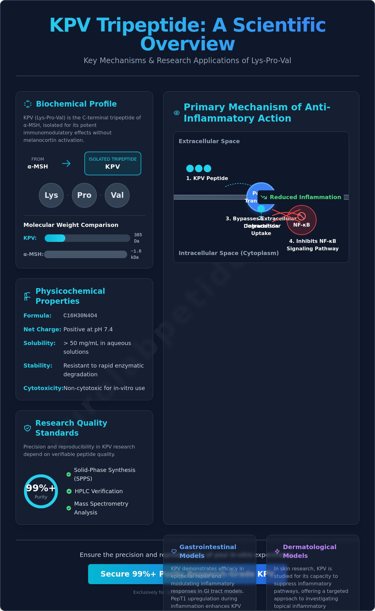
The KPV tripeptide (Lys-Pro-Val) represents the C-terminal sequence of alpha-Melanocyte-Stimulating Hormone (α-MSH). Its utility in kpv peptide anti-inflammatory research is primarily derived from its truncated structure, which lacks the His-Phe-Arg-Trp melanocortin core. This structural omission is significant. It ensures that the tripeptide doesn’t induce melanogenesis or pigmentary changes during laboratory investigation. By removing the tetrapeptide core responsible for receptor binding at MC1R, MC3R, and MC4R, researchers can isolate immunomodulatory effects without the confounding variables of systemic melanocortin activation. Within 2026 longevity and vitality research, KPV serves as a specialized tool for examining the attenuation of inflammatory signaling at the cellular level.
Origin and Synthesis from Alpha-MSH
The α-MSH neuropeptide is an evolutionary conserved molecule, with its C-terminal tripeptide sequence remaining consistent across diverse mammalian species. This conservation suggests a fundamental biological utility that persists even when the molecule is truncated. From a technical perspective, the molecular weight of KPV is approximately 385 Da, which is considerably lower than the ~1.6 kDa weight of full-length α-MSH. This reduction in mass facilitates superior cellular permeability and more efficient interaction with specific transport proteins. To ensure experimental reproducibility, solid-phase peptide synthesis (SPPS) is utilized to achieve 99%+ sequence fidelity. Every research-grade batch must undergo rigorous verification through High-Performance Liquid Chromatography (HPLC) and Mass Spectrometry to confirm the precise Lysine-Proline-Valine sequence and the absence of truncated impurities.
Physicochemical Properties and Solubility
The chemical behavior of KPV is dictated by the side chains of its constituent amino acids. The presence of Lysine at the N-terminus provides a basic character and a positive net charge at physiological pH. Proline introduces structural rigidity, while Valine adds a degree of hydrophobicity at the C-terminus. Despite the hydrophobic nature of Valine, the tripeptide maintains a high polar surface area. This results in excellent solubility parameters in aqueous environments. In laboratory settings, the peptide demonstrates stability in both sterile water and bacteriostatic saline, though its isoelectric point must be considered when formulating complex buffers for in-vitro assays. KPV is a non-cytotoxic tripeptide with high aqueous solubility for in-vitro use. Its stability profile is summarized below:
- Net Charge: Positive at pH 7.4.
- Solubility: Exceeds 50 mg/mL in aqueous solutions.
- Molecular Formula: C16H30N4O4.
- Stability: Resistant to rapid enzymatic degradation compared to full-length α-MSH.
The absence of complex secondary structures makes KPV less susceptible to denaturation than larger proteins. This inherent stability is a primary reason why it remains a centerpiece in recovery, muscle tissue, and inflammation research. Researchers prioritize the molecule because it offers a predictable chemical baseline for high-stakes precision modeling.
Primary Mechanisms of Anti-Inflammatory Action in Laboratory Models
The functional efficacy of KPV is largely dependent on its specialized transport mechanism rather than surface receptor binding. In kpv peptide anti-inflammatory research, the molecule’s interaction with the PepT1 (SLC15A1) transporter is identified as the primary gateway for its biological activity. Unlike larger peptides that require complex endocytic processes, KPV utilizes this di/tripeptide transporter to achieve rapid intracellular concentrations. This direct access allows the tripeptide to bypass extracellular degradation and interact directly with cytoplasmic signaling complexes.
PepT1-Mediated Intracellular Transport
PepT1 is a high-capacity, low-affinity proton-coupled transporter primarily expressed in the apical membrane of the small intestine. While its expression is typically low in the healthy colon, significant induction occurs during inflammatory states. Research indicates that exposure to pro-inflammatory stimuli, such as TNF-α, upregulates PepT1 expression in colonic epithelial cells. This creates a feedback loop where the presence of inflammation facilitates the uptake of the very molecule intended to modulate it. Detailed studies on KPV uptake and intestinal inflammation demonstrate that this transport is more efficient for the KPV tripeptide than for many dipeptide analogs. This is likely due to its specific lysine-proline-valine sequence. This mechanism isn’t restricted to intestinal tissue; PepT1 expression has also been documented in extra-intestinal sites, including certain immune cells, which broadens the scope of KPV’s potential research applications.
Downregulation of NF-κB and Cytokine Secretion
Once KPV achieves intracellular access, it exerts a potent inhibitory effect on the NF-κB signaling pathway. This transcription factor is a central regulator of the inflammatory response, controlling the expression of genes involved in cytokine production. KPV interferes with the nuclear translocation of the p65 RelA subunit. By blocking this translocation, the peptide prevents the assembly of the transcription complex required for the synthesis of pro-inflammatory cytokines. This molecular blockade is a verifiable metric for assessing the compound’s anti-inflammatory potential.
Quantitative analysis in LPS-stimulated macrophage models shows a marked reduction in the secretion of TNF-α, IL-1β, and IL-6. This suppression is often accompanied by a decrease in myeloperoxidase (MPO) activity, which serves as a clinical indicator for neutrophil infiltration and oxidative stress in inflamed tissue models. Beyond NF-κB, KPV interacts with secondary transcription factors like AP-1, suggesting a multi-targeted approach to suppressing cellular stress. For those conducting recovery, muscle tissue, and inflammation research, these data points provide a standardized framework for measuring KPV’s impact on systemic and localized inflammatory markers. The precision of these interactions underscores why KPV is a prioritized compound for high-stakes biochemical modeling.
Comparative Analysis of KPV Efficacy Across Tissue-Specific Research Models
The utility of kpv peptide anti-inflammatory research is most evident when examining its performance across specialized tissue models. While the biochemical pathway remains consistent, the phenotypic outcomes vary significantly between gastrointestinal, dermatological, and respiratory systems. Researchers utilize these models to determine how the tripeptide interacts with tissue-specific stressors and barrier functions. By benchmarking KPV against other compounds, such as BPC-157 in tissue repair models, a more nuanced understanding of its niche within the recovery research stack emerges. This comparative approach is essential for establishing standardized protocols in recovery, muscle tissue, and inflammation research.
Intestinal Barrier and Gut Health Studies
In gastrointestinal research, KPV is frequently investigated for its ability to maintain intestinal barrier integrity. Studies utilizing Caco-2 cell lines demonstrate that the tripeptide modulates the expression of critical tight junction proteins, specifically Occludin and Zonula Occludens-1 (ZO-1). This modulation is essential for preventing the paracellular transport of luminal antigens. Quantitative assessments of KPV’s anti-inflammatory mechanisms show a significant reduction in IL-8 secretion, which is a primary chemoattractant for neutrophils. By suppressing these signals at the gut microbiota-immune interface, KPV helps stabilize the epithelial environment under chemically induced stress. It remains a primary target for researchers focused on chronic inflammatory bowel models where epithelial repair is the objective metric of success.
Dermatological and Bronchial Epithelial Models
The scope of KPV research extends into respiratory and dermatological applications, where epithelial surfaces are exposed to environmental pathogens. In 16HBE14o- bronchial epithelial cells, the tripeptide has shown the capacity to suppress the release of pro-inflammatory mediators like IL-8 and eotaxin. This suggests a potential for modulating airway inflammation in laboratory settings. KPV functions as a dual-action agent, exhibiting both potent anti-inflammatory properties and distinct antimicrobial modulation within laboratory models. This dual capacity is particularly relevant in dermatological research, where KPV is studied for its role in suppressing inflammation induced by antimicrobial peptides like LL-37. Unlike BPC-157, which is often prioritized for its angiogenic and direct tissue-healing properties, KPV is valued for its specific regulation of the cytokine cascade and its non-receptor-mediated intracellular access. This technical distinction allows for high-stakes precision when designing multi-compound research protocols for immune system research.
Methodological Considerations for KPV Peptide Stability and Reconstitution
The integrity of kpv peptide anti-inflammatory research is contingent upon the maintenance of molecular stability throughout the experimental lifecycle. Because the Lys-Pro-Val tripeptide is susceptible to environmental degradation, standardized handling protocols are required to ensure reproducible data. Deviations in storage temperature or reconstitution technique often lead to peptide denaturation, which compromises the accuracy of in-vitro signaling assays. Precision in these early stages is a non-negotiable standard for professional laboratory inquiry.
Lyophilized KPV powder must be stored at -20°C for long-term stability. While short-term benchtop exposure is generally tolerated, extended storage at room temperature increases the risk of oxidation and proteolytic cleavage. When preparing the compound for application, investigators should utilize a peptide reconstitution calculator to determine exact molarity based on the specific vial mass. Mechanical agitation, such as high-speed vortexing, is strictly contraindicated as it may induce peptide shearing. Instead, the diluent should be added along the side of the vial, followed by gentle manual inversion to achieve a homogenous solution.
Handling and Storage Protocols
The shelf-life of reconstituted KPV is significantly shorter than its lyophilized form. Once in solution, the peptide should be maintained at 4°C and utilized within 7 to 14 days. For research requiring longer durations, the solution must be divided into single-use aliquots and frozen at -20°C or -80°C. This strategy eliminates the need for repeated freeze-thaw cycles, which are known to degrade the primary amino acid sequence. Additionally, KPV demonstrates sensitivity to light and pH fluctuations. Using sterile, buffered solutions like phosphate-buffered saline (PBS) helps maintain the tripeptide at a physiological pH, ensuring its net charge remains consistent for transport studies.
Purity and Quality Verification
Quality is treated as a verifiable metric rather than a subjective claim. High-Performance Liquid Chromatography (HPLC) is the industry standard for determining the purity percentage of research-grade KPV. A purity level of 99% or higher is required to ensure that observed biological effects are attributable to the KPV molecule and not to residual reagents or truncated sequences. Furthermore, Mass Spectrometry (MS) is employed to verify the exact molecular weight and sequence fidelity of the Lys-Pro-Val chain. In high-stakes research environments, batches are also screened for endotoxin levels. Low endotoxin counts are critical to prevent non-specific immune activation, which could otherwise confound results in recovery, muscle tissue, and inflammation research. Standardizing these analytical benchmarks allows for the absolute security of the experimental data.
Quality Standards for KPV in In-Vitro Research Applications
The integrity of kpv peptide anti-inflammatory research is fundamentally linked to the purity of the chemical reagents utilized. In laboratory settings; even minor impurities can trigger non-specific cellular responses, leading to false-positive data or skewed signaling interpretations. EuroLab Peptides implements a multi-level quality control process that begins with sequence-verified synthesis and concludes with final batch validation. Central to this process is the utilization of third-party tested peptides, which provides an objective layer of verification beyond internal manufacturing reports. This commitment to external validation ensures that each vial meets the rigorous demands of the specialized scientific community.
Sourcing High-Purity KPV in Europe
Professional investigators must navigate the complexities of buying research peptides safely in a market often characterized by inconsistent quality benchmarks. Every batch of KPV is accompanied by a Certificate of Analysis (COA), which documents the results of HPLC and Mass Spectrometry testing. These certificates verify that the peptide maintains a purity level of 99% or greater. Localized European distribution in 2026 offers significant logistical advantages, particularly regarding cold-chain delivery. By maintaining a regional supply chain, the risks of temperature fluctuations during transit are mitigated, preserving the molecular stability discussed in previous sections. All products are strictly designated with ‘Research Use Only’ (RUO) labeling, ensuring compliance with regional regulatory standards for chemical synthesis and distribution.
Conclusion: KPV as a Tool for Inflammation Research
KPV remains a unique tool for studying non-receptor-mediated melanocortin effects due to its PepT1-targeted intracellular transport. Its capacity to modulate the NF-κB pathway without the confounding effects of systemic melanocortin activation makes it an essential compound for kpv peptide anti-inflammatory research. The technical review provided here underscores the importance of precise handling, tissue-specific modeling, and high-purity sourcing. This tripeptide offers a standardized mechanism for investigating epithelial repair and cytokine suppression across multiple research models.
It’s critical to reiterate that these compounds are intended solely for laboratory research purposes. They’re not for human consumption, and no medical advice is provided or implied. Adherence to these safety protocols is a non-negotiable standard for professional inquiry. To ensure the success of your next experimental protocol, you can View KPV Technical Specifications at EuroLab Peptides and access detailed analytical data for this tripeptide.
Advancing the Precision of Melanocortin Signaling Studies
The technical landscape of kpv peptide anti-inflammatory research is defined by the requirement for molecular precision and verifiable purity. By leveraging the PepT1 transporter; this tripeptide provides a reliable model for studying intracellular cytokine suppression without the pigmentary variables of traditional melanocortins. Maintaining standardized storage at -20°C and utilizing precise reconstitution methods are critical for preserving the sequence fidelity of the Lysine-Proline-Valine chain. These methodological safeguards ensure that experimental observations remain accurate and reproducible across diverse tissue-specific models.
EuroLab Peptides facilitates these high-stakes investigations by providing third-party lab tested compounds with 99%+ purity and secure European-based logistics. Our products adhere strictly to Research Use Only (RUO) standards and aren’t intended for human consumption or therapeutic use. We invite you to Secure High-Purity KPV for Your 2026 Research Portfolio to ensure the integrity of your laboratory findings. The data generated from these models will continue to refine our collective understanding of epithelial barrier integrity and systemic recovery mechanisms. Professional inquiry depends on the quality of the tools utilized; we provide the foundation for that success.
Frequently Asked Questions
What is the primary mechanism of action for KPV peptide in research?
The primary mechanism of action for KPV involves the inhibition of the NF-κB signaling pathway following PepT1-mediated intracellular translocation. This interaction prevents the nuclear translocation of the p65 subunit, effectively downregulating the transcription of pro-inflammatory cytokines such as TNF-α and IL-6. This specific pathway is a core focus in kpv peptide anti-inflammatory research due to its ability to modulate immune responses without activating systemic melanocortin receptors.
Is KPV peptide derived from a natural hormone?
KPV is a C-terminal tripeptide fragment derived from the naturally occurring alpha-Melanocyte-Stimulating Hormone (α-MSH). While α-MSH is a 13-amino acid neuropeptide, KPV consists only of the Lysine-Proline-Valine sequence. This structural truncation allows the molecule to retain the immunomodulatory properties of the parent hormone while eliminating the tetrapeptide core responsible for inducing melanogenesis in laboratory models.
How should KPV be stored to maintain its anti-inflammatory properties?
Lyophilized KPV powder should be stored at -20°C to ensure long-term stability and prevent proteolytic degradation. Once the peptide is reconstituted, it must be refrigerated at 4°C and utilized within a 14-day window to maintain its full biological activity. For experiments requiring extended timelines, investigators should aliquot the solution and store it at -80°C to minimize the structural damage caused by repeated freeze-thaw cycles.
What is the role of the PepT1 transporter in KPV uptake?
The PepT1 (SLC15A1) transporter acts as the primary gateway for KPV’s entry into the cytoplasm of epithelial and immune cells. This proton-coupled transporter is often upregulated during inflammatory states, which facilitates the targeted uptake of KPV in stressed tissue models. This mechanism is distinct from receptor-mediated endocytosis, allowing for more rapid and efficient intracellular concentrations of the tripeptide within the experimental environment.
Can KPV be used in combination with other peptides like BPC-157 in studies?
KPV is frequently utilized alongside other compounds like BPC-157 within specialized recovery research stacks to evaluate synergistic effects on epithelial repair and barrier function. While BPC-157 is often studied for its angiogenic and growth factor modulation, KPV provides a complementary focus on the direct suppression of the cytokine cascade. This combination is a standard approach in recovery, muscle tissue, and inflammation research models.
What purity level is required for KPV in peer-reviewed research?
A purity level of 99% or higher is the non-negotiable standard for KPV intended for peer-reviewed research and high-stakes in-vitro modeling. This benchmark is verified through High-Performance Liquid Chromatography (HPLC) and Mass Spectrometry (MS) to confirm sequence fidelity and the absence of truncated impurities. Using peptides with lower purity levels risks introducing confounding variables that can invalidate experimental data and compromise reproducibility.
How does KPV differ from full-length alpha-MSH in laboratory models?
KPV differs from full-length alpha-MSH primarily through its lower molecular weight and the absence of the His-Phe-Arg-Trp melanocortin core. At approximately 385 Da, KPV possesses superior cellular permeability compared to the 1.6 kDa parent molecule. Furthermore, because it lacks the core binding sequence, KPV doesn’t activate MC1R receptors; this allows researchers to study anti-inflammatory effects without triggering pigmentary changes in laboratory models.
What are the common solvents used for KPV reconstitution?
Common solvents for KPV reconstitution include sterile water or bacteriostatic saline, depending on the requirements of the specific in-vitro assay. Phosphate-buffered saline (PBS) is also frequently employed to maintain a stable physiological pH, which is essential for transport studies involving the PepT1 gateway. In kpv peptide anti-inflammatory research, the choice of solvent must account for both the solubility of the tripeptide and the sensitivity of the cellular model.
