

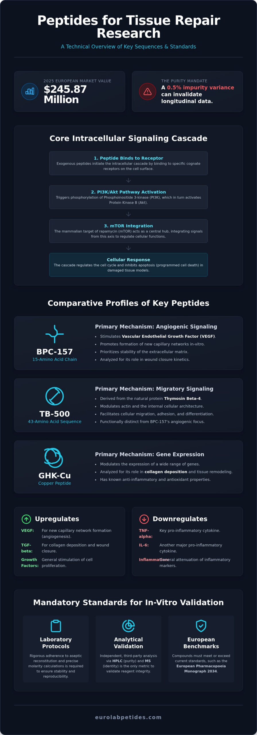
In the 2025 European peptide market, valued at USD 245.87 million, the selection of peptides for tissue repair research requires a margin of error so narrow that even a 0.5% variance in impurity can invalidate months of longitudinal data. For the professional investigator, the ambiguity surrounding synthetic sequences often represents a primary barrier to assay reproducibility. It’s understood that the integrity of any signaling pathway study is fundamentally limited by the analytical purity of the reagent. Without transparent third-party data, the risk of off-target effects or inconsistent cellular responses remains unacceptably high.
This analysis provides a technical evaluation of peptide-mediated cellular regeneration and the rigorous standards mandated for in-vitro validation. We’ll identify specific sequences for targeted signaling pathways and establish a reliable protocol for reconstitution and stability. The discussion also clarifies the current 2026 European benchmarks, including the European Pharmacopoeia Monograph 2034 standards, to ensure investigators can source verified, high-purity compounds for serious professional inquiry.
Key Takeaways
- Analyze the intracellular signaling pathways, including PI3K and mTOR, that facilitate the molecular mechanisms of peptide-mediated cellular regeneration.
- Distinguish between the angiogenic properties of BPC-157 and the migratory signaling of TB-500 to optimize the selection of peptides for tissue repair research.
- Examine the role of GHK-Cu in gene expression modulation and the impact of growth hormone secretagogues on metabolic signaling within in-vitro research models.
- Adopt rigorous laboratory protocols for aseptic reconstitution and precise molarity calculations to ensure the stability and reproducibility of synthetic sequences.
- Validate reagent integrity by identifying the benchmarks for research-grade purity and the necessity of independent third-party HPLC and MS analysis.
The Molecular Landscape of Peptide-Mediated Tissue Repair
Peptides are defined as short-chain amino acid sequences, typically consisting of 2 to 100 residues. In laboratory environments, these molecules function as critical biochemical messengers that initiate and regulate complex cellular responses. Unlike endogenous repair mechanisms, which are often limited by localized physiological constraints, exogenous peptide modulation allows for the targeted activation of specific intracellular cascades. The efficacy of peptides for tissue repair research depends entirely on the precision of these molecular interactions. Synthetic sequences like BPC-157 are frequently utilized to observe how specific chains influence the extracellular matrix and cellular integrity.
The primary pathways involved in cellular medicine include the Phosphoinositide 3-kinase (PI3K), Protein Kinase B (Akt), and the mammalian target of rapamycin (mTOR) signaling networks. This axis acts as a central hub for integrating signals from growth factors. When a research-grade peptide binds to its cognate receptor, it triggers the phosphorylation of PI3K, which subsequently activates Akt. This cascade is essential for regulating the cell cycle and inhibiting apoptosis in damaged tissue models. High-purity reagents are non-negotiable in this field. Even minor contaminants can disrupt the delicate equilibrium of an in-vitro assay, leading to skewed results that can’t be replicated. Analytical validation through High-Performance Liquid Chromatography (HPLC) is the only metric that ensures the stability of these molecular interactions.
Signaling Pathways in Cellular Regeneration
The upregulation of growth factors, specifically Vascular Endothelial Growth Factor (VEGF) and Transforming Growth Factor-beta (TGF-beta), remains a focal point in regeneration studies. Peptides often modulate these factors to stimulate angiogenesis and fibroblast activity. VEGF modulation is critical for studying the formation of new capillary networks in-vitro, while TGF-beta is analyzed for its role in collagen deposition and wound closure kinetics. Additionally, the attenuation of inflammatory markers through cytokine regulation is a key metric. Monitoring the reduction of pro-inflammatory cytokines, such as TNF-alpha and IL-6, allows researchers to quantify the anti-inflammatory efficacy of specific peptide sequences. Research indicates that peptide concentration rates directly correlate with cellular proliferation, but these rates must be meticulously controlled within the laboratory environment to avoid cytotoxic effects.
Distinguishing In-Vitro Models from Clinical Outcomes
In-vitro models provide a controlled environment to observe cellular migration, adhesion, and differentiation, yet they possess inherent limitations. They don’t fully replicate the systemic complexity of a living organism. Therefore, the scope of current peptides for tissue repair research is strictly confined to laboratory development and in-vitro analysis. Strict adherence to research-only guidelines is mandatory for all laboratory personnel. These investigations prioritize the identification of molecular first principles over clinical predictions. By focusing on how synthetic sequences influence isolated cell lines, researchers establish the foundational data necessary for understanding regenerative signaling without the confounding variables of systemic physiology.
Mechanistic Profiles of BPC-157 and TB-500 in Recovery Models
The structural divergence between BPC-157 and TB-500 dictates their specific utility in laboratory assays. BPC-157 is characterized as a 15-amino acid pentadecapeptide, while TB-500 is a 43-amino acid sequence derived from Thymosin Beta-4. These differences in molecular weight and sequence length require distinct analytical validation. The verification of sequence identity via Mass Spectrometry (MS) is a fundamental requirement to ensure that peptides for tissue repair research meet the necessary purity thresholds. Without this verification, the distinction between angiogenic and migratory mechanisms remains speculative, compromising the integrity of in-vitro data.
While these compounds are often grouped together, their molecular targets are functionally distinct. BPC-157 prioritizes the stability of the extracellular matrix through angiogenic signaling, whereas TB-500 modulates the internal architecture of the cell. Understanding these specialized roles is essential for researchers aiming to isolate specific regenerative variables. For a broader perspective on the biochemical utility of these molecules, reviewing Peptides: What to Know provides a baseline for understanding their function in scientific inquiry.
BPC-157: Gastric Pentadecapeptide and Angiogenic Modulation
The primary mechanism of BPC 157 involves the upregulation of Vascular Endothelial Growth Factor Receptor 2 (VEGFR2) and early growth response-1 (EGR-1). This signaling pathway is critical for facilitating fibroblast infiltration and collagen reorganization within in-vitro models. By enhancing the expression of these markers, BPC-157 acts as a potent modulator of the angiogenic response. Research models frequently utilize this pentadecapeptide to observe the acceleration of granulation tissue formation, providing a controlled environment to study the stabilization of damaged tissue structures without systemic interference.
TB-500 (Thymosin Beta-4): Actin Sequestration and Cell Migration
TB-500 operates through G-actin sequestration, a process that directly influences cytoskeletal remodeling. By binding to monomeric actin, TB-500 prevents polymerization, maintaining a pool of actin available for rapid cell migration. This mechanism is particularly relevant in dermal and corneal repair models where cellular mobility is the primary metric of interest. Additionally, TB-500 modulates matrix metalloproteinases (MMPs), which are enzymes responsible for degrading extracellular matrix components during tissue remodeling. This dual action on migration and matrix degradation makes it a unique mediator in recovery research stacks. Researchers investigating these combined pathways often utilize specific Recovery Research Stacks to observe multi-modal interactions in complex cellular assays.
Exploring the Role of GHK-Cu and Growth Hormone Secretagogues
GHK-Cu represents a distinct class of tripeptide-copper complexes that function primarily through gene expression modulation and extracellular matrix (ECM) remodeling. While the previously discussed BPC-157 and TB-500 focus on angiogenesis and actin-based migration, GHK-Cu is utilized in peptides for tissue repair research to observe the stimulation of glycosaminoglycan synthesis and the regulation of collagen deposition. The high affinity of the Gly-His-Lys sequence for copper (II) ions facilitates the delivery of this essential trace element to cellular sites, which is a critical variable in maintaining biochemical stability during in-vitro assays.
The bio-availability of copper-peptide complexes depends on the stability of the aqueous solution. Copper ions can be susceptible to oxidation or precipitation if the pH is not meticulously controlled. In laboratory settings, these complexes are often handled with specific buffers to prevent the dissociation of the copper ion from the tripeptide carrier. This precision ensures that the observed effects on cellular medicine are attributable to the intact complex rather than isolated components.
GHK-Cu: Copper Binding and Extracellular Matrix Remodeling
Research models demonstrate that GHK-Cu modulates the synthesis of both collagen type I and type III. A specific gap in current literature often fails to distinguish the concentration-dependent effects on these collagen types. GHK-Cu promotes a balanced ratio of these proteins, which is essential for physiological tissue integrity rather than disorganized fibrotic deposition. Additionally, the tripeptide exhibits anti-inflammatory effects by suppressing pro-inflammatory cytokines, specifically TNF-alpha and IL-6. This suppression is quantified in research models to determine the peptide’s capacity to attenuate inflammatory markers during the initial stages of cellular repair.
Secretagogues in Cellular Metabolism and IGF-1 Signaling
Growth hormone secretagogues (GHS), such as Ipamorelin and CJC-1295, provide a different mechanistic approach by targeting the GH/IGF-1 axis. Ipamorelin acts as a selective ghrelin receptor agonist. It’s noted for its ability to stimulate growth hormone secretion without causing a concomitant elevation in cortisol or prolactin levels. For investigators, the technical profile of Ipamorelin is defined by this high receptor selectivity, which minimizes off-target metabolic effects. CJC-1295, often utilized in conjunction with GHS, functions as a growth hormone-releasing hormone (GHRH) analog. It’s engineered to extend the half-life of GHRH, allowing for sustained signaling. Together, these molecules are analyzed for their role in muscle tissue repair research, where IGF-1 signaling is the primary driver of satellite cell activation and protein synthesis.
Laboratory Protocols: Reconstitution, Stability, and Handling
The precision of in-vitro assays is fundamentally dependent on the maintenance of peptide structural integrity post-synthesis. While the molecular mechanisms of peptides for tissue repair research are robust, the physical stability of these sequences is highly sensitive to environmental variables. Investigators must utilize strict aseptic techniques during reconstitution to prevent microbial contamination, which can introduce proteolytic enzymes that degrade the peptide chain. Precise molarity calculations are required for reproducible cellular medicine research, ensuring that the concentration of the reagent matches the intended experimental parameters.
Standardized handling procedures are essential to mitigate the risk of sequence degradation. Mechanical stress, such as vigorous agitation, and uncontrolled temperature fluctuations can lead to the denaturation of the peptide’s secondary structure. By adhering to rigorous laboratory standards, researchers ensure that the biochemical signals observed in their assays are a direct result of the peptide’s intended molecular interaction rather than artifacts of degradation.
Reconstitution Strategies for Lyophilized Peptides
The choice of diluent is a primary factor in maintaining long-term stability. Bacteriostatic water, containing 0.9% benzyl alcohol, is typically utilized to prevent bacterial growth in multi-use vials. However, for specific in-vitro cell cultures, sterile saline (0.9% NaCl) may be preferred to maintain osmotic balance. The dissolution process must be handled with high-stakes precision; the diluent should be added slowly along the side of the vial wall. Mechanical stress, such as vigorous shaking or vortexing, must be avoided as it can lead to the denaturation of complex sequences like TB-500. Gentle swirling is the standardized method for achieving complete dissolution without compromising the secondary structure of the peptide.
Storage Parameters and Stability Degradation
Peptides are most stable in their lyophilized, desiccated state. In this form, they can be stored at -20°C for up to 24 months or at -80°C for extended durations exceeding five years. Once reconstituted, the stability window narrows significantly. Reconstituted solutions should be stored at 2-8°C and used within 7 to 14 days, depending on the specific sequence’s susceptibility to hydrolysis. Repeated freeze-thaw cycles are a major source of sequence integrity loss and must be avoided. It’s recommended to aliquot the solution into single-use volumes immediately after reconstitution. Visual indicators of degradation include:
- Formation of visible precipitates or cloudiness in the solution.
- Changes in color, indicating oxidation or chemical alteration.
- Inconsistent results in bio-availability assays.
Maintaining these rigorous laboratory standards is essential for the validation of research-grade compounds. For investigators seeking high-purity reagents backed by independent third-party analysis, sourcing from the Tissue Repair & Recovery Research category ensures access to verified synthetic sequences for serious professional inquiry.
Sourcing Research-Grade Peptides in the European Market
The acquisition of peptides for tissue repair research in the European market is governed by rigorous analytical benchmarks that distinguish research-grade reagents from lower-tier alternatives. In 2026, the accepted standard for research-grade purity is $ge$ 99% as verified by High-Performance Liquid Chromatography (HPLC). A purity of $ge$ 98% is considered the absolute minimum for laboratory classification. These metrics aren’t arbitrary; they align with the European Pharmacopoeia (Ph. Eur.) Monograph 2034, which requires reporting impurities above 0.1% and qualifying them above 1.0%. For the professional user, these percentages represent a verifiable metric of quality that ensures assay reproducibility and minimizes off-target biochemical variables.
Regional logistics play a critical role in maintaining the security of the European supply chain. Given the prevalence of counterfeit products in the global market, sourcing from entities that prioritize regional regulatory compliance is a non-negotiable standard. EuroLab Peptides operates within this framework, providing European-based logistics that mitigate the risks associated with international transit and inconsistent manufacturing origins. This localized approach ensures that the high-stakes precision required for in-vitro analysis isn’t compromised by supply chain volatility.
Third-Party Analytical Validation: HPLC and MS Analysis
Independent third-party laboratory testing is the cornerstone of radical transparency in chemical synthesis. HPLC is utilized to provide a quantitative assessment of purity by separating the peptide from any residual solvents or truncated sequences. Complementary to this, Mass Spectrometry (MS) is employed to confirm the molecular weight and sequence identity, ensuring the synthetic chain matches the intended design. A Certificate of Analysis (CoA) must accompany every batch, providing researchers with the empirical data required to validate their reagents. Interpreting a CoA involves verifying the peak area on the HPLC chromatogram and ensuring the MS parent peak aligns with the theoretical molecular mass of the peptide.
EuroLab Peptides: Multi-Level Quality Protocol
EuroLab Peptides implements a multi-level quality control protocol that integrates both in-house and independent laboratory testing cycles. This proof-first approach ensures that every compound meets the rigorous demands of the specialized community. Products are strictly intended for in-vitro research and laboratory development use only; they aren’t intended for human consumption and no medical advice is provided. By prominently featuring empirical results and formal certifications, the brand establishes a persona of accountability in the chemical synthesis space. Before initiating any procurement, investigators should consult the final checklist to buy research peptides safely, ensuring that all sourcing aligns with established laboratory standards and regional compliance.
Advancing Precision in Regenerative Signaling Studies
Transitioning from theoretical molecular modeling to reproducible in-vitro data requires a synthesis of mechanistic understanding and rigorous laboratory standards. We’ve examined how the distinct pathways of BPC-157 and TB-500, alongside the gene expression modulation of GHK-Cu, provide a multi-modal framework for investigating cellular medicine. The validity of these observations remains entirely dependent on the analytical purity of the reagent. For peptides for tissue repair research, the adoption of $ge$ 99% HPLC-verified standards represents the definitive benchmark for professional inquiry in 2026.
EuroLab Peptides facilitates this high-stakes precision by providing strictly research-grade reagents backed by independent third-party HPLC and MS validation. Our European-based logistics ensure that localized regulatory compliance and supply chain security are maintained. Establishing a reliable foundation for cellular research starts with verified chemistry. We invite you to Secure High-Purity Peptides for Your Research at EuroLab Peptides and elevate your laboratory protocols with compounds engineered for absolute accuracy.
Frequently Asked Questions
What is the most effective peptide for muscle tissue repair research?
Ipamorelin and CJC-1295 are frequently utilized as the most effective agents for muscle tissue repair research due to their targeted activation of the GH/IGF-1 axis. These secretagogues facilitate the study of satellite cell activation and protein synthesis in-vitro. By maintaining a highly selective receptor affinity, they allow investigators to isolate specific metabolic variables without the confounding effects of elevated cortisol or prolactin levels during cellular assays.
How do BPC-157 and TB-500 differ in their molecular mechanisms?
BPC-157 and TB-500 differ primarily in their molecular targets; BPC-157 upregulates VEGFR2 to promote angiogenesis, while TB-500 utilizes G-actin sequestration to facilitate cellular migration. These distinct mechanisms determine their application in recovery models. BPC-157 is analyzed for its role in stabilizing the extracellular matrix, whereas TB-500 is the preferred reagent for observing cytoskeletal remodeling and matrix metalloproteinase modulation.
Can GHK-Cu peptides be used for in-vitro collagen synthesis studies?
GHK-Cu peptides are established tools for in-vitro collagen synthesis studies because they modulate the ratio between collagen type I and type III. This tripeptide-copper complex facilitates the delivery of essential trace elements to cellular sites, which is a critical variable in gene expression modulation. Researchers utilize these complexes to observe the stimulation of glycosaminoglycan synthesis and the regulation of fibrotic vs. healthy tissue deposition.
What is the recommended purity for peptides used in cellular assays?
The recommended purity for peptides for tissue repair research is a minimum of $ge$ 98%, though $ge$ 99% is the preferred benchmark for professional laboratory assays. This high threshold ensures that impurities, which must be reported if they exceed 0.1% according to Monograph 2034, don’t interfere with cellular signaling pathways. Utilizing lower-purity reagents risks the introduction of truncated sequences that can invalidate assay reproducibility.
How should lyophilized peptides for tissue repair be stored for long-term stability?
Lyophilized peptides should be stored at -20°C for short-term requirements or -80°C for long-term stability exceeding 24 months. These temperature parameters prevent chemical degradation and maintain the structural integrity of the synthetic sequence. Vials must be kept desiccated and away from light. Once reconstituted, the stability window narrows to 7 to 14 days when stored under refrigeration at 2-8°C.
Why is third-party testing critical for research-grade peptides?
Third-party testing is critical because it provides an objective, independent validation of HPLC purity and Mass Spectrometry sequence identity. This external verification ensures that the reagent’s chemical profile matches its technical specifications. For investigators, this transparency is essential for mitigating the risks associated with inconsistent reagent quality, which is a primary cause of failure in in-vitro tissue repair research.
Are peptides for tissue repair research intended for human use?
No, peptides intended for laboratory research are strictly prohibited from human consumption and are for in-vitro use only. These compounds are manufactured as research-grade reagents for laboratory development and chemical analysis. They don’t meet the regulatory requirements for pharmaceuticals or medicines. Any use outside of a controlled laboratory environment violates established safety standards and the intended purpose of the material.
What diluents are appropriate for reconstituting tissue repair peptides?
Appropriate diluents include bacteriostatic water (0.9% benzyl alcohol) for multi-use stability or sterile saline (0.9% NaCl) for osmotic balance in cell culture. The choice of diluent depends on the specific requirements of the in-vitro model. Bacteriostatic water is effective for preventing microbial growth, while saline is often selected when the benzyl alcohol concentration might interfere with delicate cellular medicine assays.
